ENT & Head - Neck Surgery
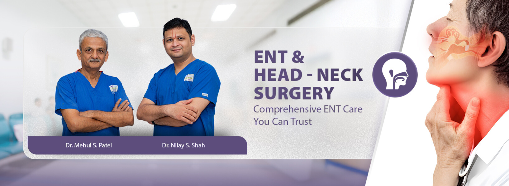
ENT & Head - Neck Surgery | IRIS Hospital
Monday to Saturdays
10:00AM to 1:00PM
5:00PM to 7:00PM
Phone: 02692-288235 (by Appointment)
Tonsil-Adenoid Coblation Surgery
- In children with Snoring, Mouth breathing and recurrent throat pain, Enlarged Tonsil and Adenoid tissue are the main causes.
- Untreated enlarged tonsil and adenoids can cause ear drum and hearing related complications, Abnormal development of face (Adenoid face), abnormal dentition and side effects of long-time antibiotics.
- In Charotar-Anand region, we are first tintroduce bloodless and painless Tonsil-Adenoid surgery for children and adults. For children with chronic tonsil infections, mouth breathing, snoring, and obstructive sleep apnoea (OSA).
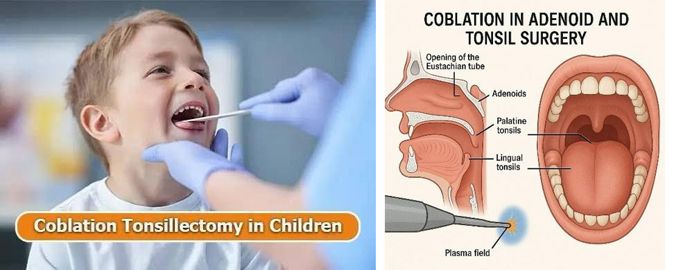
- We use latest coblation plasma technology for Tonsil-Adenoid surgery. It has obvious advantages over
conventional tonsil-adenoid surgery, such as, minimum pain, minimum bleeding, minimum length of stay and least scarring. Adenoidectomy is performed with coblation and endoscopic technology rather than conventional blind method. The obvious advantages are,
1) No bleeding,
2) No pain,
3) Complete removal- Nchance of regrowth.
Advantages of Coblation Surgery
- Coblator machine runs on principle of plasma technology which does the dissection with minimum temperature, less tissue heating, least scarring, and early resumption to work\school.
- In our hospital, coblation assisted tonsil-adenoid surgery are covered under all reputed insurance companies in cashless manner.
કાકડા અને મસાની કોબ્લેશન સર્જરી
- નસકોરા બોલવા, મોંથી શ્વાસ લેવો અને વારંવાર ગળામાં દુખાવો થતો હોય તેવા બાળકોમાં કાકડા (Tonsils) અને મસ (Adenoids) નું વધવું એ મુખ્ય કારણ છે.
- વધેલા કાકડા અને મસની સમયસર સારવાર ન કરવામાં આવે તો કાનના પડદા અને સાંભળવાની ક્ષમતાને લગતી સમસ્યાઓ, ચહેરાના અસામાન્ય વિકાસ (Adenoid Face), દાંતની અસામાન્ય રચના અને લાંબા સમય સુધી એન્ટિબાયોટિક્સ લેવાની આડઅસર થઈ શકે છે.
- ચરોતર-આણંદ વિસ્તારમાં બાળકો અને પુખ્ત વયના લોકોમાં રક્તહીન અને પીડારહિત કાકડા-મસની સર્જરી શરૂ કરનાર અમે પ્રથમ છીએ.
- વારંવાર ગળામાં ચેપ, મોંથી શ્વાસ લેવો, નસકોરા બોલવા અને Obstructive Sleep Apnoea (OSA) જેવી સમસ્યાઓ ધરાવતા બાળકો માટે આ સર્જરી અત્યંત લાભદાયક છે.
- અમે કાકડા અને મસની સર્જરી માટે અદ્યતન Coblation Plasma Technology નો ઉપયોગ કરીએ છીએ. પરંપરાગત સર્જરીની સરખામણીએ તેમાં ઓછો દુખાવો, ઓછું લોહી પડવું, ઓછો હોસ્પિટલમાં રોકાણ સમય અને ઓછા ડાઘ રહે છે.
- મસ (Adenoid) ની સર્જરી Coblation અને Endoscopic Technology દ્વારા કરવામાં આવે છે, જેથી સંપૂર્ણ નિરાકરણ મળે છે અને ફરીથી વધવાની શક્યતા ઓછી રહે છે.
કોબ્લેશન સર્જરીના ફાયદા
- કોબ્લેટર મશીન Plasma Technology ના સિદ્ધાંત પર કાર્ય કરે છે.
- ઓછા તાપમાને સર્જરી થવાથી આસપાસના ટિશ્યૂને ઓછું નુકસાન થાય છે.
- ઓછા ડાઘ રહે છે અને દર્દી ઝડપથી સામાન્ય જીવનમાં પાછા ફરી શકે છે.
- ઓછું લોહી પડે છે.
- ઓછો દુખાવો થાય છે.
- મસનું સંપૂર્ણ નિરાકરણ થાય છે અને ફરી વધવાની શક્યતા ઓછી રહે છે.
- અમારી હોસ્પિટલમાં Coblation Assisted Tonsil-Adenoid Surgery ઘણી જાણીતી ઇન્સ્યોરન્સ કંપનીઓ હેઠળ Cashless સુવિધા સાથે ઉપલબ્ધ છે.
Endoscopic Sinus & Allergy Surgery
- In our family and neighbourhood, we often see persons having frequent symptoms like excessive sneezing, watering, nasal congestion and headache.
- People usually ignore the symptoms and take frequent anti-cold over-the-counter medications.
- In frequent and long duration cases these symptoms point towards serious sinus and allergy disease for which accurate diagnosis is very important.
What is Sinus?
Paranasal sinuses are hollow cavities surrounding the nose with the function of filtering and humidifying air going into the lungs.
What is Nasal Allergy?
It is a hypersensitivity to environmental substances with which the body reacts in an unusual manner causing sneezing, watering and nasal congestion.
What is Deviated Nasal Septum (DNS)?
Nasal septum is a central partition that divides two nostrils.
Due to trauma, excessive bone growth or hereditary conditions, the nasal septum may become deviated to one or both sides.
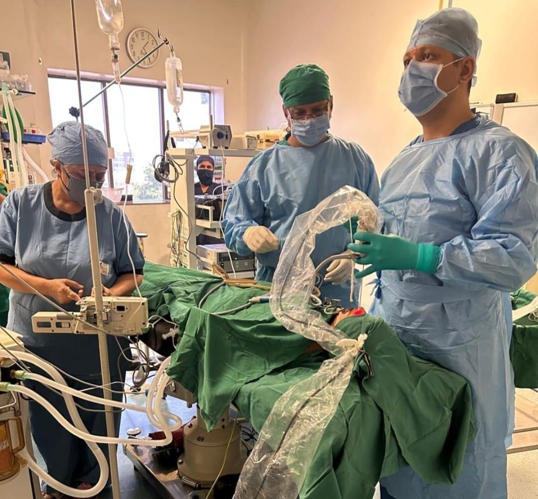
Diagnostic Facilities
- Nasal Endoscopy
- CT Scan Of Sinuses
- Allergy Skin Prick Test (SPT)
- Serum IgE
- AEC
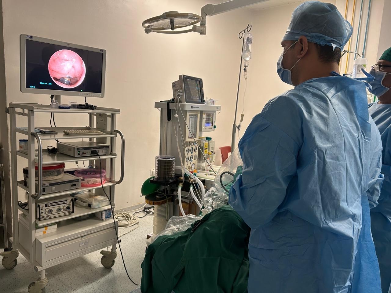
Advantages of Endoscopic Sinus Surgery
- No External Scar
- Excellent Visualization
- Precision Surgery
- No Nasal Packing
Posterior Nasal Neurectomy (PNN)
- We are among few centers in India who offer Posterior Nasal Neurectomy (PNN) Surgery.
- PNN Surgery along with sinus and septum surgery provides long term relief from excessive sneezing, runny nose and allergy symptoms.
- At IRIS Hospital we combine Sinus Surgery, Septoplasty and PNN Surgery to provide comprehensive relief.
એન્ડોસ્કોપિક સાઇનસ અને એલર્જી સર્જરી
- આપણા પરિવાર અને આસપાસમાં આપણે વારંવાર એવા લોકોને જોઈએ છીએ જેમને સતત છીંકો આવવી, નાકમાંથી પાણી પડવું, નાક બંધ થઈ જવું અને માથાનો દુખાવો જેવી તકલીફો રહેતી હોય છે.
- ઘણા લોકો આ લક્ષણોને અવગણે છે અને પોતાની રીતે શરદીની દવાઓ લેતા રહે છે.
- જો આ તકલીફો લાંબા સમય સુધી રહે તો તે ગંભીર સાઇનસ અથવા એલર્જીનો સંકેત હોઈ શકે છે. તેથી યોગ્ય નિદાન ખૂબ જ જરૂરી છે.
સાઇનસ શું છે?
પેરાનેઝલ સાઇનસ નાકની આસપાસ આવેલી પોલાણવાળી જગ્યાઓ છે જે હવાને ગાળવા, ભેજયુક્ત બનાવવાનું અને અવાજને યોગ્ય પ્રતિધ્વનિ આપવાનું કાર્ય કરે છે.
નાકની એલર્જી શું છે?
પર્યાવરણમાં રહેલા ધૂળ, પરાગકણ, ફૂગ અને અન્ય એલર્જન પ્રત્યે શરીરની અતિસંવેદનશીલ પ્રતિક્રિયાને નાકની એલર્જી કહેવામાં આવે છે.
તેના સામાન્ય લક્ષણોમાં છીંકો, નાકમાંથી પાણી પડવું, ખંજવાળ અને નાક બંધ થવું સામેલ છે.
DNS શું છે?
નાકનો પડદો (Septum) નાકને બે ભાગમાં વહેંચે છે.
જો આ પડદો વાંકો થઈ જાય તો શ્વાસ લેવામાં તકલીફ, નાક બંધ રહેવું અને સાઇનસની સમસ્યાઓ થઈ શકે છે.
નિદાન સુવિધાઓ
- નેઝલ એન્ડોસ્કોપી (Nasal Endoscopy)
- CT Scan Of Sinuses
- Allergy Skin Prick Test (SPT)
- Serum IgE
- AEC
એન્ડોસ્કોપિક સાઇનસ સર્જરીના ફાયદા
- બહાર કોઈ ડાઘ રહેતા નથી.
- સર્જરી દરમિયાન ઉત્તમ દૃશ્યતા મળે છે.
- વધુ ચોક્કસ અને સલામત સર્જરી શક્ય બને છે.
- નાકમાં પેકિંગ કરવાની જરૂરિયાત ઓછી રહે છે.
Posterior Nasal Neurectomy (PNN) Surgery
- ભારતમાં ખૂબ ઓછા કેન્દ્રો Posterior Nasal Neurectomy (PNN) Surgery ની સુવિધા આપે છે.
- PNN Surgery દ્વારા સતત છીંકો આવવી, નાકમાંથી પાણી પડવું અને એલર્જીના લક્ષણોમાં લાંબા ગાળાનો લાભ મળી શકે છે.
- IRIS Hospital માં Sinus Surgery, Septoplasty અને PNN Surgery નું સંયુક્ત આયોજન કરીને દર્દીને વધુ અસરકારક સારવાર આપવામાં આવે છે.

Sleep Apnea & Snoring Diagnosis & Treatment
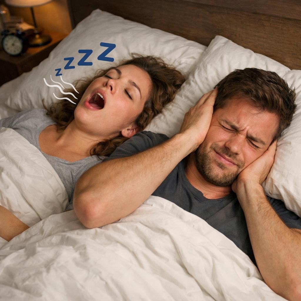
- Sleep Apnea is a serious disorder where breathing repeatedly stops during sleep.
- Apart from snoring, patients may suffer from excessive daytime sleepiness, morning fatigue, poor concentration, night-time awakening and choking during sleep.
- Many patients remain undiagnosed for years and continue to suffer from poor quality sleep.
- Sleep Apnea not only affects quality of sleep but can also lead to serious medical complications if left untreated.
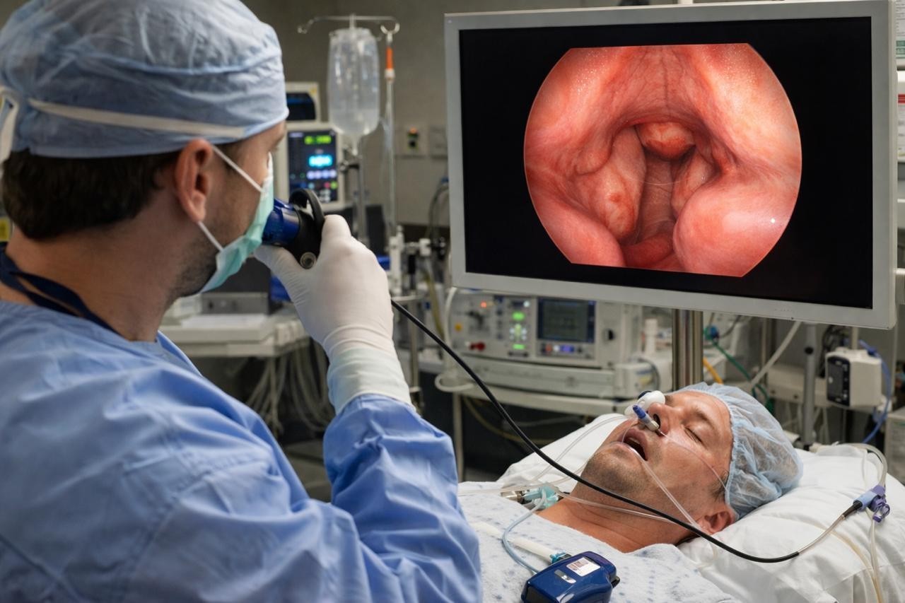
Common Symptoms
- Loud Snoring
- Excessive Daytime Sleepiness
- Morning Fatigue
- Poor Concentration
- Night-Time Awakening
- Choking During Sleep
- Irritability
- Memory Problems
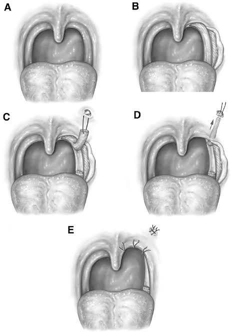
Possible Complications
- Heart Attack
- High Blood Pressure
- Brain Stroke
- Diabetes
- Memory Loss
- Poor Work Performance
- Road Traffic Accidents Due To Daytime Sleepiness
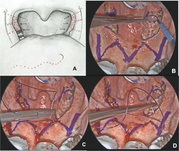
Diagnostic Facilities
- BMI Assessment
- Level-2 Sleep Study
- Drug Induced Sleep Endoscopy (DISE)
- Clinical Evaluation
- Airway Assessment
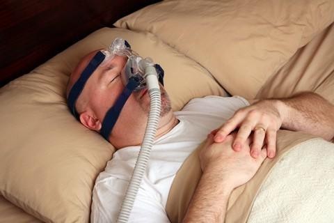
Sleep Study Classification (AHI)
- 0 – 5 : Normal
- 5 – 15 : Mild Sleep Apnea
- 15 – 30 : Moderate Sleep Apnea
- Above 30 : Severe Sleep Apnea
Treatment Options
- Weight Reduction
- Lifestyle Modification
- CPAP Therapy
- Sleep Apnea Surgery
- Medical Management
Advanced Sleep Apnea Surgeries Available
- Coblation Tonsillectomy
- Coblation Adenoidectomy
- Lingual Tonsillectomy
- Expansion Sphincter Pharyngoplasty (ESP)
- Barbed Reposition Pharyngoplasty (BRP)
- Tongue Base Reduction Surgery
- These surgeries are planned according to Sleep Study and DISE findings.
સ્લીપ એપ્નિયા અને નસકોરાની સારવાર
- સ્લીપ એપ્નિયા એ એક ગંભીર તકલીફ છે જેમાં ઊંઘ દરમિયાન વારંવાર શ્વાસ બંધ થઈ જાય છે.
- નસકોરા ઉપરાંત દર્દીને દિવસ દરમિયાન ઊંઘ આવવી, સવારમાં થાક લાગવો, ધ્યાન કેન્દ્રિત કરવામાં તકલીફ અને ઊંઘ દરમિયાન વારંવાર જાગી જવાની સમસ્યા થઈ શકે છે.
- ઘણા દર્દીઓ વર્ષો સુધી યોગ્ય નિદાન વિના આ સમસ્યાનો સામનો કરતા રહે છે.
- સમયસર સારવાર ન કરવામાં આવે તો સ્લીપ એપ્નિયા અનેક ગંભીર આરોગ્ય સમસ્યાઓનું કારણ બની શકે છે.
સામાન્ય લક્ષણો
- મોટા અવાજે નસકોરા બોલવા
- દિવસ દરમિયાન ઊંઘ આવવી
- સવારમાં થાક લાગવો
- ધ્યાન કેન્દ્રિત કરવામાં તકલીફ
- ઊંઘ દરમિયાન વારંવાર જાગી જવું
- ઊંઘ દરમિયાન ગૂંગળામણ અનુભવવી
- ચીડિયાપણું
- યાદશક્તિમાં ઘટાડો
શક્ય ગંભીર અસરો
- હાર્ટ એટેક
- હાઈ બ્લડ પ્રેશર
- બ્રેઈન સ્ટ્રોક
- ડાયાબિટીસ
- યાદશક્તિમાં ઘટાડો
- કામ કરવાની ક્ષમતામાં ઘટાડો
- દિવસ દરમિયાન ઊંઘ આવવાના કારણે અકસ્માતનું જોખમ
નિદાન સુવિધાઓ
- BMI Assessment
- Level-2 Sleep Study
- Drug Induced Sleep Endoscopy (DISE)
- Clinical Evaluation
- Airway Assessment
Sleep Study Classification (AHI)
- 0 – 5 : સામાન્ય
- 5 – 15 : હળવો સ્લીપ એપ્નિયા
- 15 – 30 : મધ્યમ સ્લીપ એપ્નિયા
- 30 થી વધુ : ગંભીર સ્લીપ એપ્નિયા
સારવારના વિકલ્પો
- વજન ઘટાડો
- જીવનશૈલીમાં સુધારો
- CPAP Therapy
- સ્લીપ એપ્નિયા સર્જરી
- દવાઓ દ્વારા સારવાર
અદ્યતન સર્જરી વિકલ્પો
- Coblation Tonsillectomy
- Coblation Adenoidectomy
- Lingual Tonsillectomy
- Expansion Sphincter Pharyngoplasty (ESP)
- Barbed Reposition Pharyngoplasty (BRP)
- Tongue Base Reduction Surgery
- દર્દીના Sleep Study અને DISE રિપોર્ટના આધારે યોગ્ય સર્જરી પસંદ કરવામાં આવે છે.
Micro Ear Surgeries
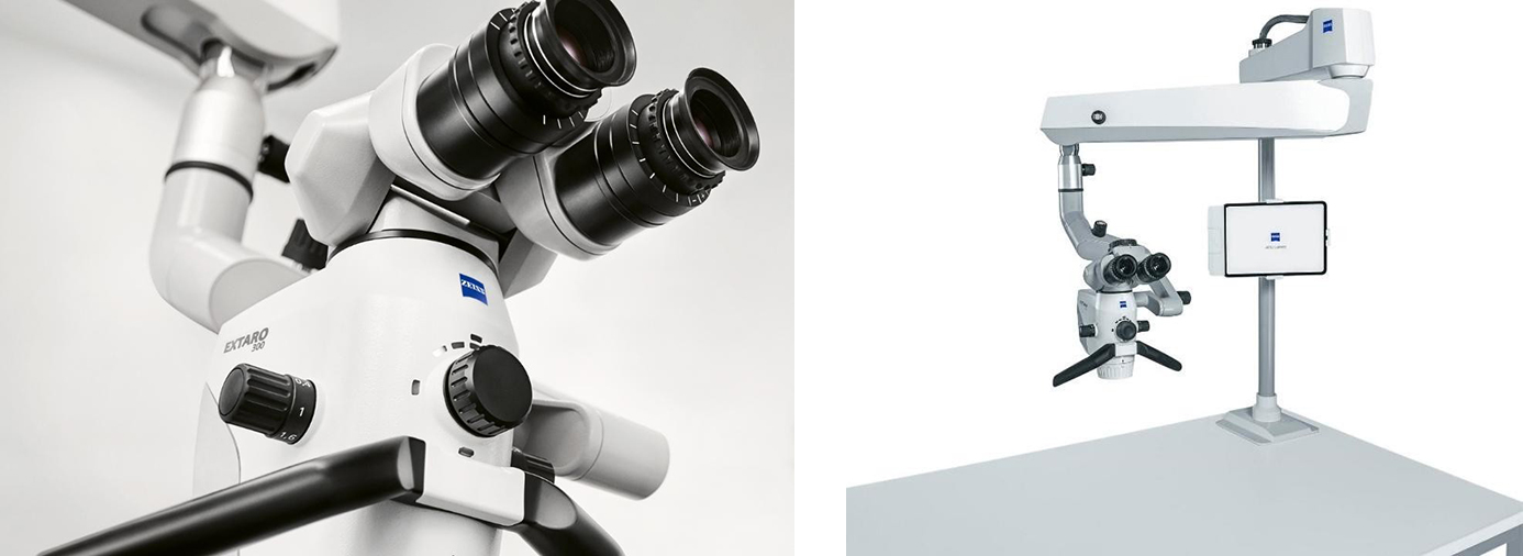
The ear is one of the most delicate and complex structures in the human body. Our ENT department specializes in Micro Ear Surgery, utilizing state-of-the-art operating microscopes to treat diseases of the middle and inner ear with pinpoint precision.
This minimally invasive approach ensures maximum preservation of healthy tissue, faster recovery times, and optimal hearing restoration.
Conditions We Treat
- Chronic Ear Infections: Persistent middle ear infections (Chronic Suppurative Otitis Media) that do not respond to medications.
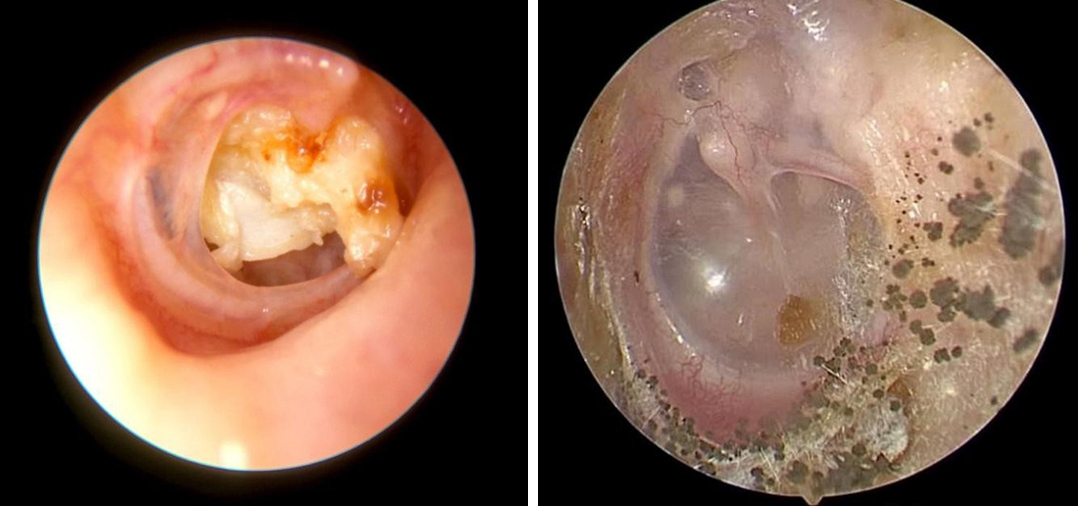
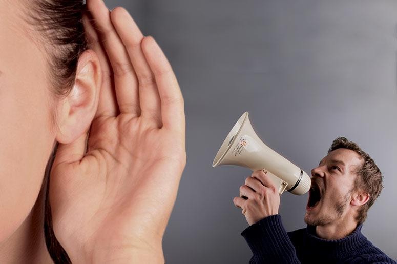
- Hearing Loss: Conductive hearing loss caused by fluid, trauma, or stiffening of the middle ear bones (Otosclerosis).
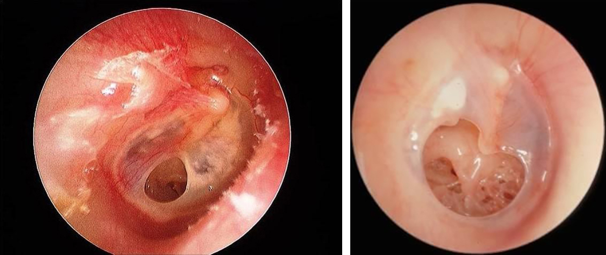
- Ruptured Eardrum: Perforations in the tympanic membrane due to injury or infection.
- Cholesteatoma: Abnormal, non-cancerous skin growths in the middle ear that can erode bone if left untreated.
- Tinnitus & Vertigo: Ringing in the ears and balance disorders linked to inner ear abnormalities.
Key Procedures Offered
- Tympanoplasty: Surgical repair of a perforated or ruptured eardrum to restore the protective barrier and improve hearing.
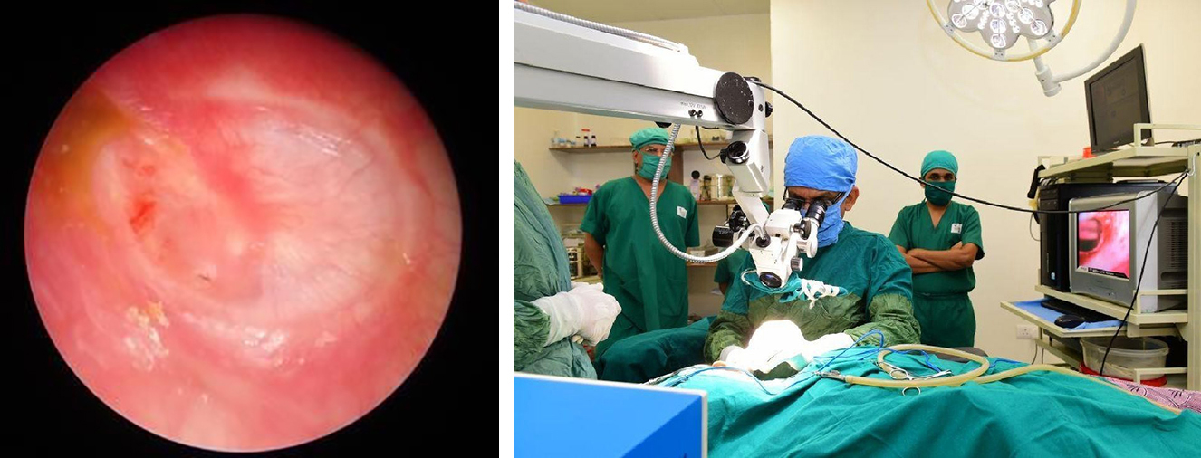
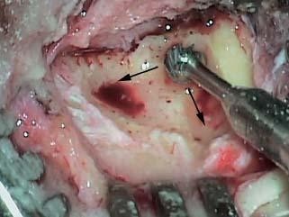
- Mastoidectomy: Removal of diseased bone cells from the mastoid (the skull bone behind the ear), commonly used to treat Cholesteatoma.
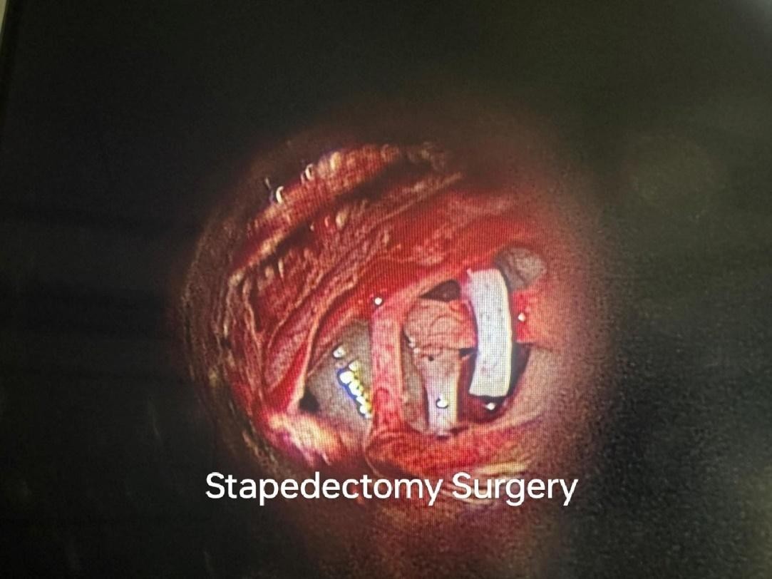
- Stapedectomy: A highly delicate procedure to treat Otosclerosis by replacing the fixed stapes bone with a micro-prosthesis, instantly restoring hearing.
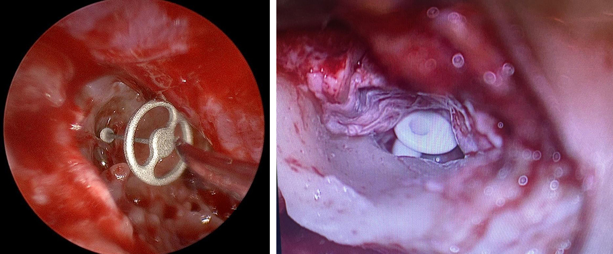
- Ossiculoplasty: Reconstruction or replacement of the tiny bones in the middle ear (malleus, incus, and stapes) to conduct sound properly.
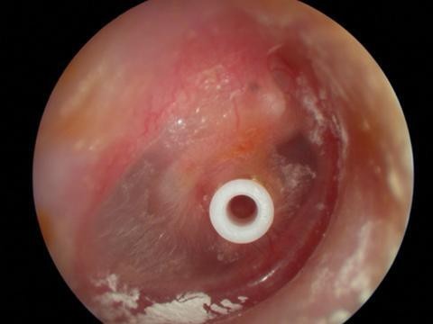
- Myringotomy & Grommet Insertion: Creating a tiny incision in the eardrum to drain fluid and inserting a microscopic tube to prevent future buildup (very common in children).
Why Choose Us for Ear Surgery?
- Advanced Optics: We use high-definition surgical microscopes that provide our surgeons with unparalleled 3D visualization of the microscopic ear structures.
- Minimally Invasive Techniques: Many procedures are done entirely through the ear canal, leaving no visible scars.
- Comprehensive Care: From pre-operative audiometry (hearing tests) to post-surgical rehabilitation, our audiology and surgical teams work seamlessly together.
કાનની માઇક્રો સર્જરી (Micro Ear Surgeries)
કાન એ માનવ શરીરની સૌથી નાજુક અને જટિલ રચનાઓમાંથી એક છે. અમારો ENT વિભાગ અત્યાધુનિક ઓપરેટિંગ માઇક્રોસ્કોપનો ઉપયોગ કરીને મધ્ય અને આંતરિક કાનના રોગોની અત્યંત સચોટ સારવાર કરવા માટે 'માઇક્રો ઇયર સર્જરી'માં વિશેષતા ધરાવે છે.
આ ન્યૂનતમ ચીરફાડવાળી પદ્ધતિ ખાતરી કરે છે કે તંદુરસ્ત પેશીઓને (tissues) ઓછામાં ઓછું નુકસાન થાય, ઝડપથી રિકવરી આવે અને સાંભળવાની ક્ષમતા વધુ સારી રીતે પાછી મેળવી શકાય.
બીમારીઓ કે જેની અમે સારવાર કરીએ છીએ
- લાંબા સમયના કાનના ચેપ: મધ્ય કાનના સતત રહેતા ચેપ (Chronic Suppurative Otitis Media - CSOM) કે જેની પર દવાની કોઈ જ અસર થતી ન હોય.
- બહેરાશ (સાંભળવામાં તકલીફ): કાનમાં પ્રવાહી જમા થવાથી, ઇજા થવાથી અથવા મધ્ય કાનના હાડકાં જકડાઈ જવાથી (ઓટોસ્ક્લેરોસિસ - Otosclerosis) આવતી બહેરાશ (Conductive Hearing Loss).
- કાનના પડદામાં કાણું (Ruptured Eardrum): ઇજા અથવા ચેપના કારણે કાનના પડદામાં થયેલું કાણું.
- કોલેસ્ટીટોમા (Cholesteatoma): મધ્ય કાનમાં ચામડીનો અસામાન્ય (કેન્સર ન હોય તેવો) વિકાસ, જેની જો સમયસર સારવાર ન કરવામાં આવે તો તે કાનના હાડકાંને નુકસાન પહોંચાડી શકે છે.
- કાનમાં અવાજ આવવો અને ચક્કર આવવા (Tinnitus & Vertigo): આંતરિક કાનની અસામાન્યતાઓને કારણે કાનમાં ગુંજવું (સીટી વાગવી) અને સંતુલન ગુમાવવાની સમસ્યાઓ.
ઉપલબ્ધ મુખ્ય સર્જિકલ પ્રક્રિયાઓ
- ટિમ્પાનોપ્લાસ્ટી (Tympanoplasty): કાનના રક્ષણાત્મક પડદાને ફરીથી બનાવવાની અને સાંભળવાની ક્ષમતા સુધારવા માટે કાણાવાળા અથવા ફાટેલા કાનના પડદાને રિપેર કરવાની સર્જરી.
- મેસ્ટોઇડેક્ટોમી (Mastoidectomy): કાનની પાછળના ખોપરીના હાડકામાંથી (મેસ્ટોઇડમાંથી) રોગગ્રસ્ત કોષોને દૂર કરવાની પ્રક્રિયા, જે સામાન્ય રીતે કોલેસ્ટીટોમાની સારવારમાં વપરાય છે.
- સ્ટેપેડેક્ટોમી (Stapedectomy): ઓટોસ્ક્લેરોસિસની સારવાર માટેની ખૂબ જ નાજુક પ્રક્રિયા, જેમાં જકડાઈ ગયેલા સ્ટેપીસ (Stapes) હાડકાને કાઢીને તેની જગ્યાએ માઇક્રો-પ્રોસ્ટેસિસ (કૃત્રિમ હાડકું) મૂકવામાં આવે છે, જેનાથી તરત જ સાંભળવાની ક્ષમતા પાછી આવે છે.
- ઓસિક્યુલોપ્લાસ્ટી (Ossiculoplasty): અવાજ યોગ્ય રીતે પસાર થાય તે માટે મધ્ય કાનના નાના હાડકાં (Malleus, Incus અને Stapes)નું પુનર્નિર્માણ અથવા તેને બદલવાની પ્રક્રિયા.
- માયરિંગોટોમી અને ગ્રોમેટ ઇન્સર્શન (Myringotomy & Grommet Insertion): પ્રવાહી બહાર કાઢવા માટે કાનના પડદામાં એક નાનો કાપો મૂકવો અને ભવિષ્યમાં ફરી પ્રવાહી જમા ન થાય તે માટે એક માઇક્રોસ્કોપિક ટ્યુબ (ગ્રોમેટ) મૂકવી (બાળકોમાં આ ખૂબ જ સામાન્ય છે).
કાનની સર્જરી માટે અમારી પસંદગી શા માટે?
- એડવાન્સ્ડ ઓપ્ટિક્સ (અત્યાધુનિક માઇક્રોસ્કોપ): અમે હાઇ-ડેફિનેશન સર્જિકલ માઇક્રોસ્કોપનો ઉપયોગ કરીએ છીએ જે અમારા સર્જનોને કાનની અતિસૂક્ષ્મ રચનાઓનું સ્પષ્ટ 3D વિઝન પ્રદાન કરે છે.
- ન્યૂનતમ ચીરફાડની તકનીકો (Minimally Invasive Techniques): ઘણી પ્રક્રિયાઓ સંપૂર્ણપણે કાનની નળી દ્વારા કરવામાં આવે છે, જેથી બહાર કોઈ જ ડાઘ દેખાતા નથી.
- સંપૂર્ણ સારવાર અને સંભાળ (Comprehensive Care): ઓપરેશન પહેલાંની ઓડિયોમેટ્રી (સાંભળવાની તપાસ)થી લઈને સર્જરી પછીના રિહેબિલિટેશન (પુનર્વસન) સુધી, અમારી ઓડિયોલોજી અને સર્જિકલ ટીમો સાથે મળીને શ્રેષ્ઠ સારવાર પૂરી પાડે છે.
Voice Box (Laryngeal) Surgery
The “voice box” or larynx contains the vocal cords, which help produce speech, protect the airway during swallowing, and assist breathing.
Voice box surgery is performed to improve voice quality, remove abnormal growths, treat airway problems, or manage early cancers of the vocal cords.
Common Conditions Treated
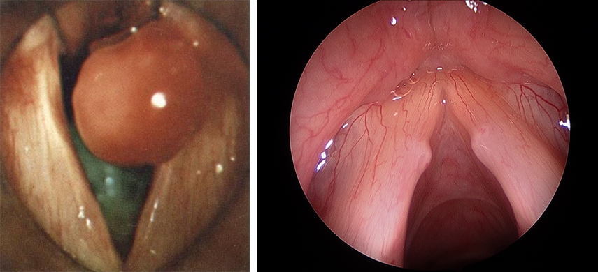
- Vocal cord polyp
- Vocal nodules
- Vocal cord cyst
- Reinke’s edema
- Vocal cord paralysis
- Hoarseness due to benign lesions
- Early vocal cord cancer
- Airway narrowing of the larynx
Common Voice Box Surgeries
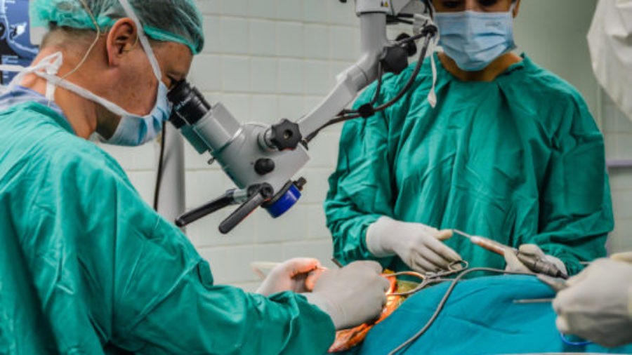
Microlaryngoscopy
A minimally invasive procedure performed through the mouth using a microscope and fine instruments.
Commonly used for:
- Polyps
- Cysts
- Nodules
- Biopsy of vocal cord lesions
Laser Vocal Cord Surgery
A precise laser technique used for selected benign lesions and early cancers.
Injection Laryngoplasty
A material is injected into the vocal cord to improve voice and swallowing in vocal cord paralysis.
Thyroplasty
An external framework surgery to improve weak or breathy voice caused by vocal cord paralysis.
Symptoms That May Require Evaluation
- Persistent hoarseness
- Voice fatigue
- Voice breaks
- Difficulty speaking loudly
- Throat discomfort while speaking
- Breathing difficulty
- Chronic cough
- Professional voice strain
Before Surgery
You may require:
- Video laryngoscopy
- Stroboscopy
- Voice assessment
- Blood tests
- Anaesthesia fitness evaluation
Smoking cessation and reflux control are strongly recommended before surgery.
What Happens During Surgery?
Most voice box surgeries are performed under general anaesthesia.
A telescope or microscope is used to visualize the vocal cords.
Fine instruments or laser are then used to treat the lesion while preserving normal vocal cord vibration.
Most patients go home the same day.
Recovery After Voice Box Surgery
Voice rest is very important.
Temporary voice rest helps proper healing of the vocal cords and reduces scarring risk.
Typical recommendations after vocal cord surgery include:
- Complete voice rest for 2–5 days in many cases.
- Avoid whispering, shouting, coughing and throat clearing.
- Gradual return to speaking under medical guidance.
- Some surgeons may advise longer restrictions depending on the procedure performed.
During Recovery
You may experience:
- Mild throat pain
- Temporary hoarseness
- Increased throat mucus
- Mild swallowing discomfort
These symptoms usually improve gradually over days to weeks.
Important Precautions After Surgery
- Do not smoke.
- Avoid alcohol and excessive caffeine.
- Stay well hydrated.
- Avoid forceful coughing or throat clearing.
- Avoid heavy lifting for a few days.
Voice Therapy
Speech and voice therapy are often essential after surgery to:
- Improve vocal quality.
- Reduce strain.
- Prevent recurrence.
- Restore professional voice performance.
When To Contact Your Doctor
Seek medical attention if you develop:
- Breathing difficulty
- Significant bleeding
- Fever
- Severe pain
- Sudden worsening of voice
- Difficulty swallowing liquids
Expected Outcomes
Most benign vocal cord surgeries provide significant improvement in:
- Hoarseness
- Vocal strength
- Voice clarity
- Vocal endurance
Recovery time varies depending on the condition treated and the extent of surgery.
These procedures are covered by all major insurance companies in cashless mode.
સ્વરપેટીની (લેરીન્જિયલ) સર્જરી
"સ્વરપેટી" અથવા લેરીંક્સમાં (Larynx) સ્વરતંતુઓ (Vocal Cords) હોય છે, જે બોલવામાં, ગળતી વખતે શ્વાસનળીનું રક્ષણ કરવામાં અને શ્વાસ લેવામાં મદદ કરે છે.
સ્વરપેટીની સર્જરી અવાજની ગુણવત્તા સુધારવા, અસામાન્ય ગાંઠ કે મસા દૂર કરવા, શ્વાસનળીની સમસ્યાઓની સારવાર કરવા અથવા સ્વરતંતુઓના પ્રારંભિક કેન્સરની સારવાર માટે કરવામાં આવે છે.
સામાન્ય બીમારીઓ કે જેની સારવાર કરવામાં આવે છે
- સ્વરતંતુના પોલિપ (મસા)
- સ્વરતંતુની ગાંઠ (નોડ્યુલ્સ)
- સ્વરતંતુની સિસ્ટ (પાણીની ગાંઠ)
- રેઇન્કેસ એડિમા (સ્વરતંતુ પર સોજો)
- સ્વરતંતુનો લકવો (પેરાલિસિસ)
- સાધારણ ગાંઠ કે મસાને કારણે અવાજ બેસી જવો
- સ્વરતંતુઓનું પ્રારંભિક કેન્સર
- સ્વરપેટીમાં શ્વાસનળી સાંકડી થવી
સ્વરપેટીની સામાન્ય સર્જરીઓ
માઇક્રોલેરીંગોસ્કોપી (Microlaryngoscopy)
માઇક્રોસ્કોપ અને બારીક સાધનોનો ઉપયોગ કરીને મોં દ્વારા કરવામાં આવતી એકદમ ઓછી ચીરફાડવાળી પ્રક્રિયા.
સામાન્ય રીતે આ માટે વપરાય છે:
- પોલિપ (મસા)
- સિસ્ટ (પાણીની ગાંઠ)
- નોડ્યુલ્સ (ગાંઠ)
- સ્વરતંતુની બીમારીના નિદાન માટે બાયોપ્સી લેવા
લેસર વોકલ કોર્ડ સર્જરી
પસંદ કરેલી સાધારણ ગાંઠો અને પ્રારંભિક કેન્સર માટે ઉપયોગમાં લેવાતી અત્યાધુનિક લેસર તકનીક.
ઇન્જેક્શન લેરીંગોપ્લાસ્ટી
સ્વરતંતુના લકવા (પેરાલિસિસ) માં અવાજ અને ગળવાની ક્ષમતા સુધારવા માટે સ્વરતંતુમાં એક પદાર્થ (દવા) ઇન્જેક્ટ કરવામાં આવે છે.
થાયરોપ્લાસ્ટી
સ્વરતંતુના લકવાને કારણે નબળા કે ધીમા પડી ગયેલા અવાજને સુધારવા માટે બહારથી ગળાના ભાગે કરવામાં આવતી સર્જરી.
લક્ષણો કે જેના માટે તપાસની જરૂર પડી શકે છે
- લાંબા સમય સુધી અવાજ બેસી જવો કે ઘોઘરો રહેવો
- બોલતી વખતે થાક લાગવો
- અવાજ તૂટી જવો
- મોટેથી બોલવામાં તકલીફ થવી
- બોલતી વખતે ગળામાં અગવડતા કે દુખાવો થવો
- શ્વાસ લેવામાં તકલીફ પડવી
- લાંબા સમયની ખાંસી
- વ્યવસાયિક કારણોસર અવાજ પર તાણ પડવું (ગાયકો, શિક્ષકો વગેરે માટે)
સર્જરી પહેલાં
તમારે નીચે મુજબની તપાસની જરૂર પડી શકે છે:
- વીડિયો લેરીંગોસ્કોપી
- સ્ટ્રોબોસ્કોપી
- અવાજની તપાસ (Voice Assessment)
- લોહીના રિપોર્ટ
- એનેસ્થેસિયા ફિટનેસ તપાસ
સર્જરી પહેલાં ધૂમ્રપાન બંધ કરવાની અને એસિડિટી / રિફ્લક્સને નિયંત્રિત કરવાની ખાસ સલાહ આપવામાં આવે છે.
સર્જરી દરમિયાન શું થાય છે?
સ્વરપેટીની મોટાભાગની સર્જરી જનરલ એનેસ્થેસિયા (સંપૂર્ણ બેભાન કરીને) હેઠળ કરવામાં આવે છે.
સ્વરતંતુઓને જોવા માટે ટેલિસ્કોપ અથવા માઇક્રોસ્કોપનો ઉપયોગ કરવામાં આવે છે.
ત્યારબાદ સ્વરતંતુના સામાન્ય કંપનને નુકસાન પહોંચાડ્યા વિના, બારીક સાધનો અથવા લેસરનો ઉપયોગ કરીને ગાંઠ/મસાની સારવાર કરવામાં આવે છે.
મોટાભાગના દર્દીઓ તે જ દિવસે ઘરે જઈ શકે છે.
સ્વરપેટીની સર્જરી પછીની રિકવરી
અવાજને આરામ આપવો (Voice Rest) ખૂબ જ મહત્વપૂર્ણ છે.
હંગામી ધોરણે અવાજને આરામ આપવાથી સ્વરતંતુઓ યોગ્ય રીતે રૂઝાય છે અને ડાઘ પડવાનું જોખમ ઘટે છે.
સ્વરતંતુની સર્જરી પછીની સામાન્ય ભલામણોમાં નીચેની બાબતોનો સમાવેશ થાય છે:
- ઘણા કિસ્સાઓમાં ૨ થી ૫ દિવસ માટે અવાજને સંપૂર્ણ આરામ આપવો.
- ધીમા અવાજે બોલવું, બૂમો પાડવી, ખાંસી ખાવી અને ગળું સાફ કરવાનું ટાળો.
- ડૉક્ટરના માર્ગદર્શન હેઠળ ધીમે ધીમે બોલવાનું શરૂ કરો.
- કરવામાં આવેલી સર્જરીના પ્રકારના આધારે કેટલાક સર્જન વધુ સમય માટે પ્રતિબંધોની સલાહ આપી શકે છે.
રિકવરી દરમિયાન
તમને નીચે મુજબનો અનુભવ થઈ શકે છે:
- ગળામાં હળવો દુખાવો
- થોડા સમય માટે અવાજ બેસી જવો
- ગળામાં કફ (મ્યુકસ) વધી જવો
- ગળવામાં હળવી અગવડતા
આ લક્ષણો સામાન્ય રીતે થોડા દિવસોથી અઠવાડિયામાં ધીમે ધીમે સુધરી જાય છે.
સર્જરી પછીની મહત્વપૂર્ણ સાવચેતીઓ
- ધૂમ્રપાન ન કરો.
- આલ્કોહોલ અને વધુ પડતા કેફીન (ચા/કોફી) નું સેવન ટાળો.
- પુષ્કળ પાણી પીવો (શરીરને હાઇડ્રેટેડ રાખો).
- જોરથી ખાંસી ખાવી કે ગળું સાફ કરવાનું ટાળો.
- થોડા દિવસો માટે ભારે વજન ઊંચકવાનું ટાળો.
વોઇસ થેરાપી (અવાજની થેરાપી)
સર્જરી પછી સ્પીચ અને વોઇસ થેરાપી ઘણીવાર નીચેના કારણોસર જરૂરી હોય છે:
- અવાજની ગુણવત્તા સુધારવા માટે.
- અવાજ પર પડતો તાણ ઘટાડવા માટે.
- સમસ્યાને ફરીથી થતી અટકાવવા માટે.
- વ્યવસાયિક અવાજનું પ્રદર્શન પુનઃસ્થાપિત કરવા માટે.
તમારા ડૉક્ટરનો સંપર્ક ક્યારે કરવો?
જો તમને નીચેનામાંથી કોઈ તકલીફ જણાય તો તરત જ ડૉક્ટરની સલાહ લો:
- શ્વાસ લેવામાં તકલીફ
- નોંધપાત્ર રક્તસ્ત્રાવ (લોહી પડવું)
- તાવ આવવો
- અસહ્ય દુખાવો થવો
- અવાજ અચાનક વધુ ખરાબ થઈ જવો
- પ્રવાહી ગળવામાં તકલીફ થવી
અપેક્ષિત પરિણામો
સાધારણ ગાંઠ માટે થતી મોટાભાગની સ્વરતંતુઓની સર્જરી નીચેની બાબતોમાં નોંધપાત્ર સુધારો લાવે છે:
- બેસી ગયેલો અવાજ
- અવાજની તાકાત (Vocal Strength)
- અવાજની સ્પષ્ટતા
- બોલવાની ક્ષમતા / ટકાઉપણું (Vocal Endurance)
સાજા થવાનો સમય (રિકવરી) બીમારીના પ્રકાર અને સર્જરી કેટલી મોટી હતી તેના પર આધાર રાખે છે.
આ પ્રક્રિયાઓ તમામ મુખ્ય ઇન્શ્યોરન્સ કંપનીઓ દ્વારા કેશલેસ સુવિધામાં આવરી લેવામાં આવે છે.
Specialized Vertigo & Balance Clinic
Experiencing vertigo—the sudden, intense sensation that you or your surroundings are spinning—can be debilitating and frightening.
Because the inner ear is the body's primary control center for balance, our dedicated Vertigo Clinic specializes in finding the exact root cause of your dizziness.
We combine advanced diagnostic technology with targeted therapies to help you regain your balance and return to your daily life with confidence.
Symptoms That Need Evaluation
- A sudden spinning or swaying sensation, especially when changing head positions.
- Unsteadiness or difficulty walking in a straight line.
- Dizziness accompanied by nausea or vomiting.
- Ringing in the ears (tinnitus), a feeling of fullness in the ear, or sudden hearing loss.
Common Conditions We Treat
- BPPV (Benign Paroxysmal Positional Vertigo): The most common cause of vertigo, triggered by tiny calcium crystals becoming dislodged in the inner ear.
- Meniere’s Disease: A chronic inner ear condition characterized by episodes of severe vertigo, fluctuating hearing loss, and ringing in the ear caused by fluid buildup.
- Vestibular Neuritis & Labyrinthitis: Inflammation of the inner ear or the vestibular nerve, usually caused by a viral infection, leading to prolonged, intense dizziness.
- Vestibular Migraines: Episodes of vertigo and sensitivity to motion that are linked to migraine conditions, sometimes without a traditional headache.
Advanced Diagnostic Services
Finding the right treatment starts with an accurate diagnosis.
Our clinic is equipped with state-of-the-art vestibular testing, allowing us to evaluate the complete balance system without relying on guesswork.
Our advanced diagnostic tools include:
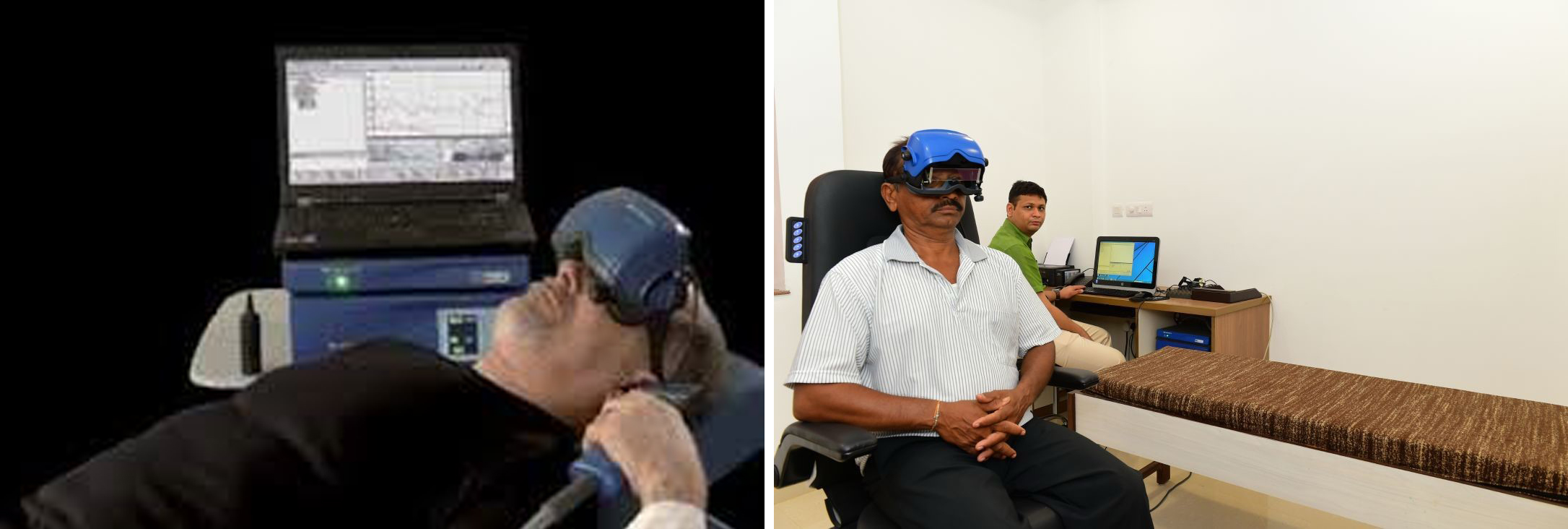
- Videonystagmography (VNG): A specialized test using infrared goggles to track precise eye movements, helping us pinpoint whether the issue lies in the inner ear or the central nervous system.
- Video Head Impulse Test (vHIT): This quick, highly advanced test uses lightweight, high-speed camera goggles to measure how well your eyes stay focused while your head is moved in different directions. It allows our specialists to independently test all six balance canals in your inner ear, detecting subtle nerve damage with high precision.
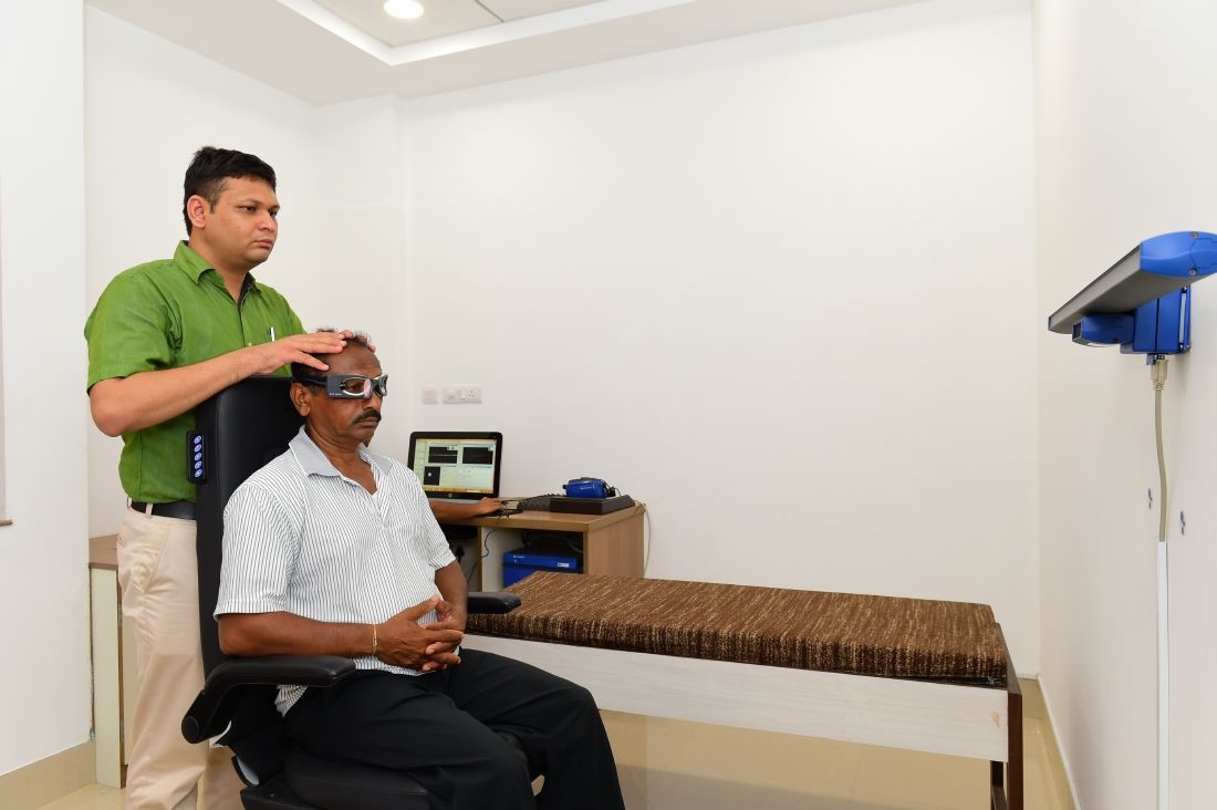
- Vestibular Evoked Myogenic Potential (VEMP): A painless test that measures how specific parts of your inner ear (the utricle and saccule) respond to sound clicks or vibrations. By placing simple sensor pads on your neck or face, VEMP helps us accurately diagnose complex conditions like Meniere's disease or thinning of the inner ear bones.
- Pure Tone Audiometry (PTA): Because hearing and balance are deeply connected, comprehensive hearing tests help us isolate specific inner ear conditions and complete your diagnostic profile.
Treatments & Therapies
- Canalith Repositioning (The Epley Maneuver): A highly effective, in-office physical maneuver that shifts dislodged crystals back to their proper place, often curing BPPV in just one or two sessions.
- Vestibular Rehabilitation Therapy (VRT): Customized, exercise-based physical therapy designed to retrain your brain to compensate for inner ear imbalances.
- Medical Management: Targeted prescription medications to control acute nausea, suppress vestibular attacks, or manage conditions like Meniere's disease.
- Intratympanic Injections: For severe, medication-resistant cases (like advanced Meniere’s), we offer minimally invasive injections of medication directly into the middle ear to calm the balance nerve.
Why Choose Our Vertigo Clinic?
- Accurate Diagnosis Over Guesswork: We don't just prescribe anti-dizziness medications to mask symptoms; we find and treat the underlying anatomical cause.
- Rapid Relief: For conditions like BPPV, our doctors can often provide immediate relief during your very first consultation.
- Multidisciplinary Approach: Our ENT surgeons work closely with audiologists and vestibular physiotherapists to provide complete, start-to-finish care.
સ્પેશિયલાઈઝ્ડ વર્ટિગો અને બેલેન્સ ક્લિનિક (ચક્કર અને સંતુલન માટેનું ખાસ ક્લિનિક)
વર્ટિગો (ચક્કર) નો અનુભવ — જેમાં અચાનક એવો તીવ્ર અહેસાસ થાય છે કે તમે અથવા તમારી આસપાસની વસ્તુઓ ગોળ ગોળ ફરી રહી છે — તે ખૂબ જ કષ્ટદાયક અને ડરામણો હોઈ શકે છે.
આંતરિક કાન એ શરીરનું સંતુલન જાળવતું મુખ્ય નિયંત્રણ કેન્દ્ર હોવાથી, અમારું ખાસ 'વર્ટિગો ક્લિનિક' તમારા ચક્કરના ચોક્કસ મૂળ કારણને શોધવામાં વિશેષતા ધરાવે છે.
તમારું સંતુલન પાછું મેળવવામાં અને આત્મવિશ્વાસ સાથે તમારા રોજિંદા જીવનમાં પાછા ફરવામાં તમને મદદ કરવા માટે અમે અત્યાધુનિક નિદાન તકનીક અને સચોટ સારવારનો સમન્વય કરીએ છીએ.
લક્ષણો કે જેના માટે તપાસની જરૂર છે
- અચાનક ચક્કર આવવા અથવા ઝોલા ખાવા જેવો અનુભવ થવો, ખાસ કરીને માથાની સ્થિતિ બદલતી વખતે.
- અસ્થિરતા લાગવી અથવા સીધી લાઇનમાં ચાલવામાં તકલીફ પડવી.
- ચક્કરની સાથે ઉબકા આવવા કે ઉલટી થવી.
- કાનમાં અવાજ આવવો (સીટી વાગવી), કાન ભારે લાગવો (બંધ થઈ ગયો હોય તેવું લાગવું), અથવા અચાનક સાંભળવાનું ઓછું થઈ જવું.
સામાન્ય બીમારીઓ કે જેની અમે સારવાર કરીએ છીએ
- BPPV (બિનાઈન પેરોક્સિસ્મલ પોઝિશનલ વર્ટિગો): ચક્કર આવવાનું આ સૌથી સામાન્ય કારણ છે, જે આંતરિક કાનમાં રહેલા કેલ્શિયમના નાના કણો (ક્રિસ્ટલ્સ) તેમની જગ્યાએથી ખસી જવાને કારણે થાય છે.
- મિનિયર્સ ડિસીઝ (Meniere’s Disease): આંતરિક કાનની આ લાંબા ગાળાની બીમારી છે, જેમાં કાનમાં પ્રવાહી ભરાવાને કારણે અચાનક સખત ચક્કર આવવા, સાંભળવામાં વધઘટ થવી અને કાનમાં અવાજ આવવાની સમસ્યા થાય છે.
- વેસ્ટિબ્યુલર ન્યુરાઇટિસ અને લેબિરિન્થાઇટિસ: આંતરિક કાન અથવા વેસ્ટિબ્યુલર નસમાં સોજો આવવો, જે સામાન્ય રીતે વાયરલ ઇન્ફેક્શનને કારણે થાય છે, જેના પરિણામે લાંબા સમય સુધી તીવ્ર ચક્કર આવે છે.
- વેસ્ટિબ્યુલર માઇગ્રેન: માઇગ્રેન (આધાશીશી) સાથે સંકળાયેલ ચક્કર આવવા અને હલનચલન પ્રત્યે સંવેદનશીલતા, જેમાં કેટલીકવાર સામાન્ય માથાનો દુખાવો ન પણ હોય.
અત્યાધુનિક નિદાન (ડાયગ્નોસ્ટિક) સેવાઓ
યોગ્ય સારવારની શરૂઆત સચોટ નિદાનથી થાય છે.
અમારું ક્લિનિક અત્યાધુનિક વેસ્ટિબ્યુલર ટેસ્ટિંગ સાધનોથી સજ્જ છે, જે અમને કોઈપણ અટકળો પર આધાર રાખ્યા વિના સંતુલનની સંપૂર્ણ સિસ્ટમનું મૂલ્યાંકન કરવાની મંજૂરી આપે છે.
અમારા અત્યાધુનિક ડાયગ્નોસ્ટિક સાધનોમાં નીચે મુજબનો સમાવેશ થાય છે:
- વીડિયોનિસ્ટેગ્મોગ્રાફી (VNG): આંખની ચોક્કસ હલનચલનને ટ્રેક કરવા માટે ઇન્ફ્રારેડ ગોગલ્સનો ઉપયોગ કરતો એક ખાસ ટેસ્ટ, જે ચક્કરની સમસ્યા આંતરિક કાનમાં છે કે સેન્ટ્રલ નર્વસ સિસ્ટમમાં (મગજમાં) તે સચોટ રીતે શોધવામાં મદદ કરે છે.
- વીડિયો હેડ ઇમ્પલ્સ ટેસ્ટ (vHIT): આ ઝડપી અને અત્યાધુનિક ટેસ્ટમાં હળવા વજનના હાઇ-સ્પીડ કેમેરાવાળા ગોગલ્સનો ઉપયોગ થાય છે. જ્યારે તમારા માથાને જુદી જુદી દિશામાં ફેરવવામાં આવે ત્યારે તમારી આંખો કેટલી સારી રીતે ફોકસ જાળવી રાખે છે તે માપવામાં આવે છે. તે અમારા નિષ્ણાતોને આંતરિક કાનની સંતુલન જાળવતી તમામ છ નળીઓનું સ્વતંત્ર રીતે પરીક્ષણ કરવાની મંજૂરી આપે છે, જે ઉચ્ચ ચોકસાઈ સાથે નસના હળવા નુકસાનને પણ શોધી કાઢે છે.
- વેસ્ટિબ્યુલર ઇવોક્ડ માયોજેનિક પોટેન્શિયલ (VEMP): આ એક પીડારહિત ટેસ્ટ છે જે માપે છે કે તમારા આંતરિક કાનના ચોક્કસ ભાગો (યુટ્રિકલ અને સેક્યુલ) અવાજ અથવા ધ્રુજારી (Vibrations) સામે કેવી પ્રતિક્રિયા આપે છે. તમારી ગરદન અથવા ચહેરા પર સામાન્ય સેન્સર પેડ મૂકીને કરવામાં આવતો VEMP ટેસ્ટ, મિનિયર્સ ડિસીઝ અથવા આંતરિક કાનના હાડકાં પાતળા થવા જેવી જટિલ પરિસ્થિતિઓનું સચોટ નિદાન કરવામાં મદદ કરે છે.
- પ્યોર ટોન ઓડિયોમેટ્રી (PTA): સાંભળવાની ક્ષમતા અને સંતુલન એકબીજા સાથે ગાઢ રીતે સંકળાયેલા હોવાથી, સાંભળવાની આ સંપૂર્ણ તપાસ અમને આંતરિક કાનની ચોક્કસ સમસ્યાઓને અલગ પાડવામાં અને તમારા નિદાનને પૂર્ણ કરવામાં મદદ કરે છે.
સારવાર અને થેરાપીઓ
- કેનાલિથ રિપોઝિશનિંગ (એપ્લે મેન્યુવર - The Epley Maneuver): ક્લિનિકમાં જ કરવામાં આવતી અત્યંત અસરકારક શારીરિક કસરત, જે જગ્યા પરથી ખસી ગયેલા ક્રિસ્ટલ્સને (કણોને) તેમની યોગ્ય જગ્યાએ પાછા મોકલે છે. આનાથી ઘણીવાર માત્ર એક કે બે સિટિંગમાં જ BPPV મટી જાય છે.
- વેસ્ટિબ્યુલર રિહેબિલિટેશન થેરાપી (VRT): આંતરિક કાનના અસંતુલનને પહોંચી વળવા માટે તમારા મગજને ફરીથી તૈયાર કરવા (ટ્રેનિંગ આપવા) માટે ખાસ બનાવાયેલી, કસરત આધારિત ફિઝિકલ થેરાપી.
- મેડિકલ મેનેજમેન્ટ (દવાઓ દ્વારા સારવાર): તીવ્ર ઉબકાને નિયંત્રિત કરવા, ચક્કરના હુમલાને અટકાવવા અથવા મિનિયર્સ ડિસીઝ જેવી પરિસ્થિતિઓને નિયંત્રિત કરવા માટે અપાતી સચોટ દવાઓ.
- ઇન્ટ્રાટિમ્પેનિક ઇન્જેક્શન (કાનના પડદામાં ઇન્જેક્શન): દવાઓથી મટતા ન હોય તેવા ગંભીર કેસો માટે (જેમ કે એડવાન્સ્ડ મિનિયર્સ ડિસીઝ), અમે સંતુલન જાળવતી નસને શાંત કરવા સીધા મધ્ય કાનમાં દવા પહોંચાડવા માટે ન્યૂનતમ ચીરફાડવાળા ઇન્જેક્શન આપીએ છીએ.
વર્ટિગો ક્લિનિક માટે અમારી પસંદગી શા માટે?
- અટકળોને બદલે સચોટ નિદાન: અમે માત્ર લક્ષણોને છુપાવવા માટે ચક્કર વિરોધી દવાઓ લખી આપતા નથી; અમે શારીરિક સંરચનાની ખામીના મૂળ કારણને શોધીએ છીએ અને તેની સારવાર કરીએ છીએ.
- ઝડપી રાહત: BPPV જેવી બીમારીઓ માટે, અમારા ડૉક્ટરો ઘણીવાર તમારી પ્રથમ મુલાકાત દરમિયાન જ તાત્કાલિક રાહત પ્રદાન કરી શકે છે.
- મલ્ટિડિસિપ્લિનરી અભિગમ (સર્વાંગી સારવાર): શરૂઆતથી અંત સુધી સંપૂર્ણ કાળજી પૂરી પાડવા માટે અમારા ENT સર્જનો ઓડિયોલોજિસ્ટ અને વેસ્ટિબ્યુલર ફિઝિયોથેરાપિસ્ટ સાથે મળીને કામ કરે છે.
Comprehensive Audiology & Cochlear Implant Center
Hearing connects us to the people we love and the world around us.
Whether you are dealing with age-related hearing loss or seeking care for a child born with profound deafness, our Audiology and Cochlear Implant Center offers a complete spectrum of care.
By combining advanced diagnostic testing with the latest in hearing aid technology and surgical implants, our team of ENT surgeons, audiologists, and speech therapists work together to help you restore the joy of sound.
Diagnostic Audiology Services
Accurate treatment begins with precise testing.
Our sound-treated audiology labs are equipped to test patients of all ages, from newborns to the elderly.
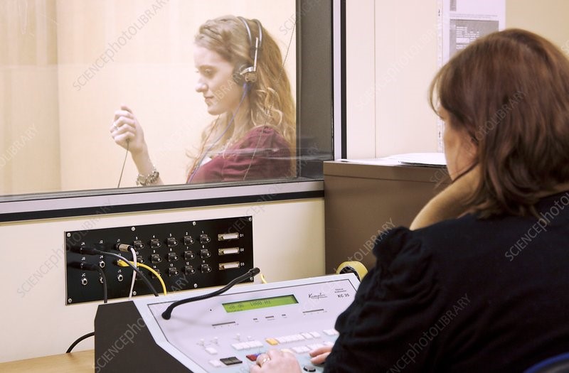
- Pure Tone Audiometry (PTA) & Tympanometry: The gold standard for assessing hearing thresholds and evaluating the health and pressure of the middle ear.
- Otoacoustic Emissions (OAE): A quick, painless screening test ideal for newborns and infants to check the function of the outer hair cells in the inner ear.
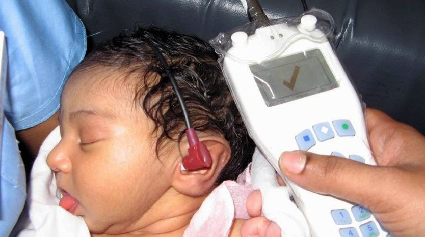
- Brainstem Evoked Response Audiometry (BERA / ABR): An advanced, objective test that measures how the hearing nerve and brain respond to sound. This is essential for evaluating hearing in infants, small children, or patients who cannot participate in standard tests.
- Auditory Steady-State Response (ASSR): Provides highly specific, frequency-by-frequency hearing threshold estimates, crucial for programming hearing aids or assessing cochlear implant candidacy in young children.
Advanced Hearing Solutions
For patients with mild to severe hearing loss, we offer comprehensive hearing rehabilitation:
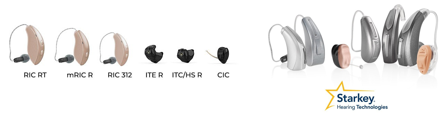
- Digital Hearing Aid Fitting: We dispense and program the latest generation of invisible, rechargeable, and Bluetooth-enabled digital hearing aids tailored to your specific hearing curve and lifestyle.
- Tinnitus Management: Specialized masking devices and sound therapies for patients experiencing ringing in the ears.
The Cochlear Implant Program
For patients who receive little or no benefit from powerful hearing aids, a cochlear implant can be a life-changing solution.
Unlike a hearing aid that simply amplifies sound, a cochlear implant is a surgically implanted device that bypasses damaged parts of the inner ear and directly stimulates the hearing nerve.
Who Can Benefit?
- Children Born with Deafness: Early intervention is critical. Implanting children at a young age allows them to develop normal speech, language, and social skills.
- Adults with Severe to Profound Hearing Loss: For those who have lost their hearing later in life and can no longer communicate effectively even with the best hearing aids.
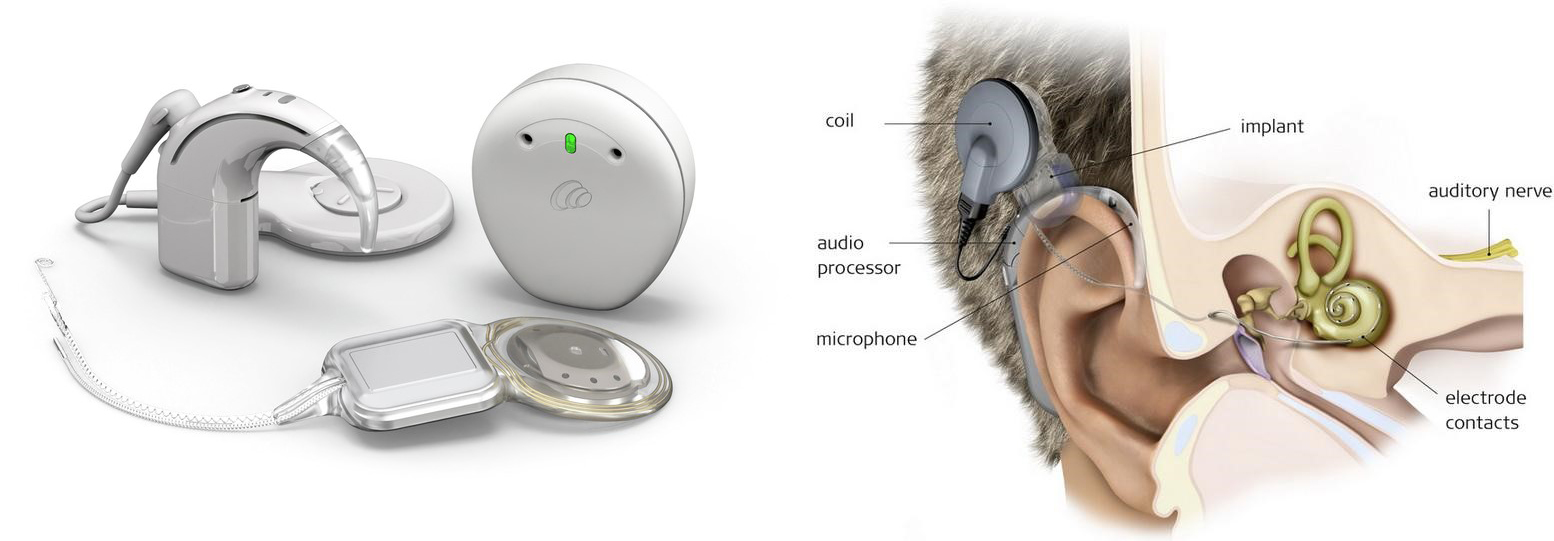
Our Complete Implant Journey
- Pre-Implant Evaluation: A thorough medical, radiological (HRCT/MRI), and audiological assessment to determine if the patient is a suitable candidate.
- Implant Surgery: Our specialized ENT surgeons perform this delicate, minimally invasive procedure to carefully place the internal electrode array into the cochlea.
- Switch-On & Mapping: A few weeks after surgery, the external processor is activated. Our audiologists carefully "map" and fine-tune the device to ensure comfortable and clear sound perception.
- Auditory Verbal Therapy (AVT): Surgery is only the first step. Our dedicated speech and language therapists work closely with patients (especially children) and their families to train the brain to understand these new electronic sound signals and develop natural speech.
Why Choose Us for Hearing Care?
- Care Under One Roof: From your first diagnostic hearing test to surgery, device mapping, and years of speech therapy, our entire multidisciplinary team is located in one facility.
- Lifelong Support: A cochlear implant is a lifelong commitment. We partner with families for the long haul, providing ongoing device upgrades, mapping, and rehabilitation support.
કોમ્પ્રિહેન્સિવ ઓડિયોલોજી એન્ડ કોકલિયર ઇમ્પ્લાન્ટ સેન્ટર (સંપૂર્ણ શ્રવણ અને કોકલિયર ઇમ્પ્લાન્ટ કેન્દ્ર)
સાંભળવાની શક્તિ આપણને આપણા પ્રિયજનો અને આપણી આસપાસની દુનિયા સાથે જોડે છે.
પછી ભલે તમે ઉંમરને સંબંધિત બહેરાશનો સામનો કરી રહ્યા હોવ અથવા જન્મથી સંપૂર્ણ બહેરાશ ધરાવતા બાળકની સારવાર માટે જોઈ રહ્યા હોવ, અમારું 'ઓડિયોલોજી અને કોકલિયર ઇમ્પ્લાન્ટ સેન્ટર' સારવારની સંપૂર્ણ સુવિધા પ્રદાન કરે છે.
અત્યાધુનિક નિદાન (ટેસ્ટિંગ), લેટેસ્ટ હિયરિંગ એડ (કાનના મશીન) ટેક્નોલોજી અને સર્જિકલ ઇમ્પ્લાન્ટ્સના સમન્વયથી, અમારા ENT સર્જનો, ઓડિયોલોજિસ્ટ અને સ્પીચ થેરાપિસ્ટની ટીમ તમને અવાજનો આનંદ પાછો મેળવવામાં મદદ કરવા માટે એકસાથે કામ કરે છે.
ઓડિયોલોજી નિદાન (ડાયગ્નોસ્ટિક) સેવાઓ
યોગ્ય સારવારની શરૂઆત સચોટ તપાસથી થાય છે.
અમારી સાઉન્ડ-પ્રૂફ ઓડિયોલોજી લેબ નવજાત શિશુથી લઈને વૃદ્ધો સુધીના તમામ ઉંમરના દર્દીઓની તપાસ કરવા માટે સજ્જ છે:
- પ્યોર ટોન ઓડિયોમેટ્રી (PTA) અને ટિમ્પેનોમેટ્રી: સાંભળવાની ક્ષમતા (બહેરાશનું પ્રમાણ) માપવા અને મધ્ય કાનના દબાણ અને સ્વાસ્થ્યની તપાસ માટેની આદર્શ અને ઉત્તમ પદ્ધતિ.
- ઓટોએકોસ્ટિક એમિશન્સ (OAE): નવજાત શિશુઓ અને નાના બાળકો માટે આદર્શ એવી આ એક ઝડપી અને પીડારહિત તપાસ છે, જે આંતરિક કાનના 'આઉટર હેર સેલ્સ' ની કામગીરી તપાસે છે.
- બ્રેઈનસ્ટેમ ઇવોક્ડ રિસ્પોન્સ ઓડિયોમેટ્રી (BERA / ABR): એક અત્યાધુનિક તપાસ જે માપે છે કે સાંભળવાની નસ અને મગજ અવાજ પ્રત્યે કેવી પ્રતિક્રિયા આપે છે. શિશુઓ, નાના બાળકો અથવા સામાન્ય તપાસમાં સહકાર ન આપી શકતા દર્દીઓમાં સાંભળવાની ક્ષમતા તપાસવા માટે આ અત્યંત જરૂરી છે.
- ઓડિટરી સ્ટેડી-સ્ટેટ રિસ્પોન્સ (ASSR): આ ફ્રીક્વન્સી અનુસાર સાંભળવાની ક્ષમતાનો ખૂબ જ સચોટ અંદાજ પૂરો પાડે છે, જે નાના બાળકોમાં હિયરિંગ એડના પ્રોગ્રામિંગ અથવા કોકલિયર ઇમ્પ્લાન્ટની યોગ્યતા નક્કી કરવા માટે ખૂબ જ મહત્વપૂર્ણ છે.
અત્યાધુનિક શ્રવણ ઉકેલો (Advanced Hearing Solutions)
હળવીથી લઈને ગંભીર બહેરાશ ધરાવતા દર્દીઓ માટે, અમે સંપૂર્ણ શ્રવણ પુનર્વસન (Hearing Rehabilitation) પ્રદાન કરીએ છીએ:
- ડિજિટલ હિયરિંગ એડ ફિટિંગ: અમે તમારી સાંભળવાની ક્ષમતા અને જીવનશૈલીને અનુરૂપ, નવી પેઢીના દેખાય નહીં તેવા (ઇનવિઝિબલ), રિચાર્જ કરી શકાય તેવા અને બ્લૂટૂથ-સક્ષમ ડિજિટલ હિયરિંગ એડ્સ (કાનના મશીન) પ્રદાન કરીએ છીએ અને તેનું પ્રોગ્રામિંગ કરીએ છીએ.
- ટિનીટસ મેનેજમેન્ટ: કાનમાં અવાજ આવવા (સીટી વાગવા) ની સમસ્યા અનુભવતા દર્દીઓ માટે ખાસ માસ્કિંગ ઉપકરણો અને સાઉન્ડ થેરાપી.
કોકલિયર ઇમ્પ્લાન્ટ પ્રોગ્રામ
જે દર્દીઓને પાવરફુલ હિયરિંગ એડ્સથી પણ ઓછો કે બિલકુલ ફાયદો થતો નથી, તેમના માટે કોકલિયર ઇમ્પ્લાન્ટ એક જીવન બદલી નાખનારો ઉકેલ બની શકે છે.
કાનના મશીન (જે માત્ર અવાજને મોટો કરે છે) થી વિપરીત, કોકલિયર ઇમ્પ્લાન્ટ એ એક સર્જરી દ્વારા બેસાડવામાં આવતું ઉપકરણ છે જે આંતરિક કાનના ક્ષતિગ્રસ્ત ભાગોને છોડીને સીધું સાંભળવાની નસને ઉત્તેજિત કરે છે.
કોને ફાયદો થઈ શકે?
- જન્મથી બહેરાશ ધરાવતા બાળકો: વહેલું નિદાન અને સારવાર ખૂબ જ મહત્વપૂર્ણ છે. નાની ઉંમરે બાળકોમાં ઇમ્પ્લાન્ટ કરવાથી તેઓમાં સામાન્ય વાણી, ભાષા અને સામાજિક કૌશલ્યોનો વિકાસ થાય છે.
- ગંભીર બહેરાશ ધરાવતા પુખ્ત વયના લોકો: જેઓ ઉંમર વધવાની સાથે સાંભળવાની શક્તિ ગુમાવી ચૂક્યા છે અને શ્રેષ્ઠ કાનના મશીન સાથે પણ યોગ્ય રીતે વાતચીત કરી શકતા નથી તેમના માટે.
અમારી સંપૂર્ણ ઇમ્પ્લાન્ટ જર્ની (પ્રક્રિયા)
- ઇમ્પ્લાન્ટ પહેલાની તપાસ (Pre-Implant Evaluation): દર્દી ઇમ્પ્લાન્ટ માટે યોગ્ય છે કે નહીં તે નક્કી કરવા માટે સંપૂર્ણ તબીબી, રેડિયોલોજિકલ (HRCT/MRI) અને ઓડિયોલોજિકલ તપાસ.
- ઇમ્પ્લાન્ટ સર્જરી: અમારા નિષ્ણાત ENT સર્જનો દ્વારા અત્યંત ચોકસાઈપૂર્વક અને ઓછામાં ઓછી ચીરફાડવાળી સર્જરી દ્વારા આંતરિક ઇલેક્ટ્રોડને કોક્લિયામાં બેસાડવામાં આવે છે.
- સ્વિચ-ઓન અને મેપિંગ: સર્જરીના થોડા અઠવાડિયા પછી, બહારના મશીનને (પ્રોસેસરને) ચાલુ કરવામાં આવે છે. અમારા ઓડિયોલોજિસ્ટ સ્પષ્ટ અને આરામદાયક અવાજ સંભળાય તે માટે ડિવાઇસને કાળજીપૂર્વક "મેપ" (સેટિંગ) કરે છે.
- ઓડિટરી વર્બલ થેરાપી (AVT): સર્જરી એ માત્ર પહેલું પગલું છે. અમારા નિષ્ણાત સ્પીચ અને લેંગ્વેજ થેરાપિસ્ટ દર્દીઓ (ખાસ કરીને બાળકો) અને તેમના પરિવારો સાથે મળીને મગજને આ નવા ઇલેક્ટ્રોનિક અવાજના સંકેતો સમજવા અને કુદરતી વાણી વિકસાવવા માટે તાલીમ આપે છે.
શ્રવણ (કાનની) સંભાળ માટે અમારી પસંદગી શા માટે?
- એક જ છત નીચે સંપૂર્ણ સારવાર: તમારી પ્રથમ તપાસથી લઈને સર્જરી, ડિવાઇસ મેપિંગ અને વર્ષોની સ્પીચ થેરાપી સુધી, અમારી સંપૂર્ણ નિષ્ણાત ટીમ એક જ જગ્યાએ ઉપલબ્ધ છે.
- આજીવન સહાય (Lifelong Support): કોકલિયર ઇમ્પ્લાન્ટ એ આજીવન કમિટમેન્ટ છે. અમે લાંબા ગાળા માટે પરિવારો સાથે જોડાઈએ છીએ, અને ડિવાઇસ અપગ્રેડેશન, મેપિંગ તેમજ પુનર્વસનમાં સતત સહાય પ્રદાન કરીએ છીએ.
Head & Neck and Skull Base Surgery
Conditions involving the head, neck, and the base of the skull require extraordinary precision. These areas are home to critical nerves, blood vessels, and structures that control your voice, swallowing, hearing, and facial expressions.
Our specialized surgical team provides expert, multidisciplinary care for both benign (non-cancerous) and malignant (cancerous) conditions.
We focus not only on completely removing the disease but also on preserving your quality of life and appearance through minimally invasive techniques.
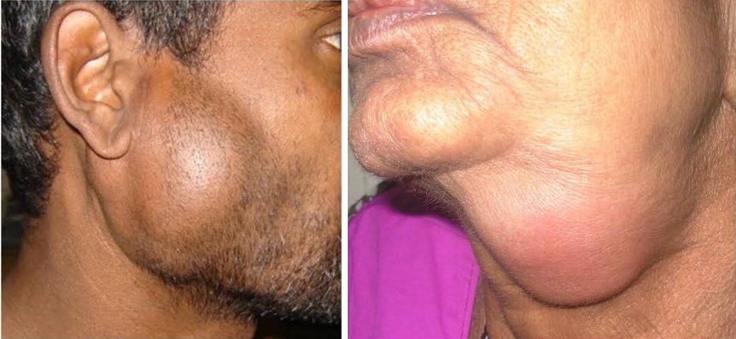
Head & Neck Conditions We Treat
We evaluate and treat a wide variety of masses, cysts, and tumors in the head and neck region, including:
- Thyroid & Parathyroid Disorders: Nodules, cysts, enlarged thyroids (goiters), and thyroid cancers.
- Salivary Gland Tumors: Lumps or recurrent infections in the parotid and submandibular glands.
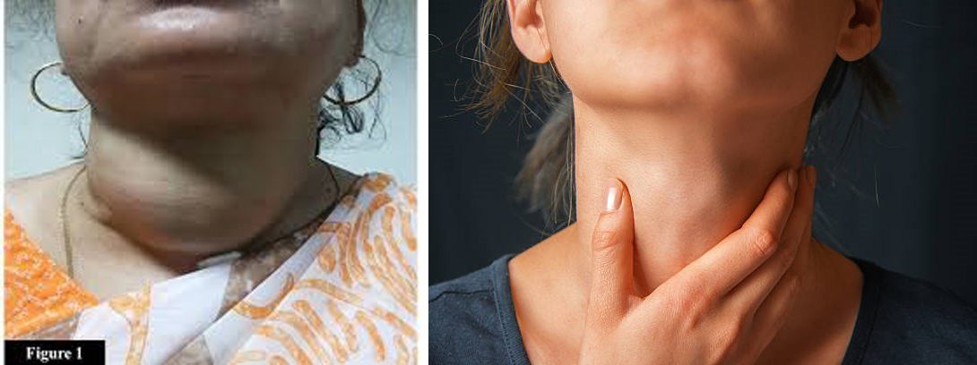
- Neck Masses & Cysts: Congenital cysts (like thyroglossal duct cysts) and unexplained lymph node enlargements.
- Throat & Voice Box (Laryngeal) Lesions: Growths on the vocal cords or surrounding tissues affecting voice and breathing.
- Oral Cavity Conditions: Benign and malignant growths of the mouth, tongue, and throat.
Skull Base Surgery: Reaching the Hard-to-Reach
The "skull base" is the deeply buried, complex area at the bottom of the skull where the brain sits, directly behind the nose and eyes.
In the past, reaching tumors here required large incisions.
Today, our ENT surgeons use the natural corridors of the nose to perform Endoscopic Endonasal Skull Base Surgery.
Common Skull Base Conditions We Treat
- CSF Leaks: Repairing tears in the brain's protective lining that cause cerebrospinal fluid to leak into the nose.
- Pituitary Tumors: Removing tumors from the pituitary gland seamlessly through the nasal passages.
- Acoustic Neuromas (Vestibular Schwannomas): Managing benign tumors on the hearing and balance nerves.
- Sinonasal Tumors: Treating complex growths that start in the sinuses and extend toward the brain or eyes.
Advanced Surgical Technology
- High-Definition Endoscopy: Using ultra-HD cameras and specialized angled instruments, we can remove deep-seated skull base tumors entirely through the nostrils—meaning no facial incisions, no visible scars, and a much faster recovery.
Why Choose Us for Complex Surgeries?
- Function-Preserving Philosophy: Our primary goal is curing the disease while meticulously preserving your ability to speak, eat, smile, and look like yourself.
- Multidisciplinary Tumor Board: Complex head, neck, and skull base cases are never treated in isolation. Our ENT surgeons collaborate closely with neurosurgeons, oncologists, and radiologists to create a comprehensive, customized treatment plan for you.
- Advanced Reconstructive Care: When large areas of tissue must be removed, our team utilizes advanced reconstructive techniques to restore natural appearance and function.
હેડ એન્ડ નેક અને સ્કલ બેઝ (માથા, ગરદન અને ખોપરીના પાયાની) સર્જરી
માથા, ગરદન અને ખોપરીના પાયાને (સ્કલ બેઝને) લગતી બીમારીઓની સારવારમાં અસાધારણ ચોકસાઈની જરૂર હોય છે.
આ વિસ્તારોમાં મહત્વપૂર્ણ નસો, રક્તવાહિનીઓ અને એવી રચનાઓ આવેલી છે જે તમારા અવાજ, ગળવાની ક્રિયા, સાંભળવાની ક્ષમતા અને ચહેરાના હાવભાવને નિયંત્રિત કરે છે.
અમારી નિષ્ણાત સર્જિકલ ટીમ સાધારણ (કેન્સર વગરની) અને જીવલેણ (કેન્સરવાળી) બંને પ્રકારની બીમારીઓ માટે નિષ્ણાત અને મલ્ટિડિસિપ્લિનરી સારવાર પ્રદાન કરે છે.
અમારું ધ્યાન માત્ર રોગને સંપૂર્ણપણે દૂર કરવા પર જ નથી, પરંતુ ન્યૂનતમ ચીરફાડવાળી તકનીકો (Minimally Invasive Techniques) દ્વારા તમારા જીવનની ગુણવત્તા અને દેખાવને જાળવી રાખવા પર પણ છે.
હેડ એન્ડ નેક (માથા અને ગરદન) ની બીમારીઓ જેની અમે સારવાર કરીએ છીએ
અમે માથા અને ગરદનના વિસ્તારમાં થતી વિવિધ પ્રકારની ગાંઠો, સિસ્ટ (પાણીની ગાંઠ) અને ટ્યુમરનું નિદાન અને સારવાર કરીએ છીએ, જેમાં નીચે મુજબનો સમાવેશ થાય છે:
- થાઇરોઇડ અને પેરાથાઇરોઇડની સમસ્યાઓ: નોડ્યુલ્સ (ગાંઠ), સિસ્ટ, થાઇરોઇડનું વધવું (ગોઇટર) અને થાઇરોઇડ કેન્સર.
- લાળગ્રંથિની ગાંઠ (Salivary Gland Tumors): પેરોટિડ અને સબમેન્ડિબ્યુલર ગ્રંથીઓમાં થતી ગાંઠ અથવા વારંવાર થતો ચેપ.
- ગરદનની ગાંઠો અને સિસ્ટ: જન્મજાત સિસ્ટ (જેમ કે થાયરોગ્લોસલ ડક્ટ સિસ્ટ) અને કારણ વગર લસિકા ગ્રંથીઓ (Lymph Nodes) નું વધવું.
- ગળા અને સ્વરપેટીની (લેરીન્જિયલ) બીમારીઓ: સ્વરતંતુઓ અથવા તેની આસપાસની પેશીઓ પર થતો અસામાન્ય વિકાસ જે અવાજ અને શ્વાસોચ્છ્વાસને અસર કરે છે.
- મુખગુહાની (Oral Cavity) બીમારીઓ: મોં, જીભ અને ગળામાં થતી સાધારણ અથવા કેન્સરની ગાંઠો.
સ્કલ બેઝ (ખોપરીના પાયાની) સર્જરી: પહોંચવામાં મુશ્કેલ જગ્યાઓ સુધી પહોંચવું
"સ્કલ બેઝ" એ ખોપરીના તળિયે આવેલો ખૂબ ઊંડો અને જટિલ વિસ્તાર છે, જ્યાં મગજ ગોઠવાયેલું છે, જે નાક અને આંખોની બરાબર પાછળ આવેલો છે.
ભૂતકાળમાં, અહીં રહેલી ગાંઠ (ટ્યુમર) સુધી પહોંચવા માટે મોટા કાપા મૂકવાની જરૂર પડતી હતી.
આજે, અમારા ENT સર્જનો એન્ડોસ્કોપિક એન્ડોનેઝલ સ્કલ બેઝ સર્જરી કરવા માટે નાકના કુદરતી રસ્તાઓનો ઉપયોગ કરે છે.
સામાન્ય સ્કલ બેઝ બીમારીઓ જેની અમે સારવાર કરીએ છીએ
- CSF લીક (મગજના પ્રવાહીનું લીકેજ): મગજના રક્ષણાત્મક પડમાં પડેલા કાણા/ચીરાને રિપેર કરવું, જેના કારણે મગજનું પ્રવાહી (સેરેબ્રોસ્પાઇનલ ફ્લુઇડ) નાકમાં લીક થાય છે.
- પિટ્યુટરી ટ્યુમર: નાકના માર્ગ દ્વારા પિટ્યુટરી ગ્રંથિમાંથી ગાંઠને સરળતાથી દૂર કરવી.
- એકોસ્ટિક ન્યુરોમાસ (વેસ્ટિબ્યુલર શ્વાનોમાસ): સાંભળવા અને સંતુલન રાખતી નસો પર થતી સાધારણ ગાંઠોની સારવાર.
- સાઇનોનેઝલ ટ્યુમર: એવી જટિલ ગાંઠોની સારવાર જે સાઇનસથી શરૂ થાય છે અને મગજ અથવા આંખો તરફ ફેલાય છે.
અત્યાધુનિક સર્જિકલ ટેક્નોલોજી
- ઇન્ટ્રાઓપરેટિવ નર્વ મોનિટરિંગ: થાઇરોઇડ, પેરોટિડ અથવા કાનની સર્જરી દરમિયાન, અમે ચહેરાની અને સ્વરતંતુની નસોને સતત ટ્રેક કરવા માટે અત્યાધુનિક નર્વ-મોનિટરિંગ સિસ્ટમનો ઉપયોગ કરીએ છીએ. આનાથી સર્જરી પછી ચહેરા પર નબળાઈ આવવાનું કે અવાજ બદલાઈ જવાનું જોખમ નોંધપાત્ર રીતે ઘટી જાય છે.
- સર્જિકલ નેવિગેશન (ઇમેજ ગાઇડન્સ સિસ્ટમ): જટિલ સાઇનસ અને સ્કલ બેઝ સર્જરીઓ માટે, અમે ઓપરેશન થિયેટરમાં 3D GPS જેવી ટ્રેકિંગ સિસ્ટમનો ઉપયોગ કરીએ છીએ. આ સિસ્ટમ સર્જનને મગજ અને આંખોથી મિલીમીટરના અંતરે પણ તેમના સાધનોને રિયલ-ટાઇમમાં ટ્રેક કરવાની મંજૂરી આપે છે, જેથી મહત્તમ સલામતી સુનિશ્ચિત થાય છે.
- હાઇ-ડેફિનેશન એન્ડોસ્કોપી: અલ્ટ્રા-HD કેમેરા અને ખાસ પ્રકારના સાધનોનો ઉપયોગ કરીને, અમે સ્કલ બેઝની ઊંડી ગાંઠોને સંપૂર્ણપણે નાક દ્વારા જ દૂર કરી શકીએ છીએ — જેનો અર્થ એ છે કે ચહેરા પર કોઈ કાપો નહીં, કોઈ ડાઘ નહીં અને દર્દીને ખૂબ જ ઝડપી રિકવરી મળે છે.
જટિલ સર્જરીઓ માટે અમારી પસંદગી શા માટે?
- કાર્ય જાળવી રાખવાની ફિલોસોફી (Function-Preserving Philosophy): અમારો મુખ્ય ઉદ્દેશ્ય રોગને મટાડવાની સાથે તમારી બોલવાની, ખાવાની, હસવાની ક્ષમતા અને તમારા મૂળ દેખાવને સાવચેતીપૂર્વક જાળવી રાખવાનો છે.
- મલ્ટિડિસિપ્લિનરી ટ્યુમર બોર્ડ: માથા, ગરદન અને સ્કલ બેઝના જટિલ કેસોની સારવાર ક્યારેય એકલા કરવામાં આવતી નથી. તમારા માટે સંપૂર્ણ અને વ્યક્તિગત સારવાર યોજના બનાવવા માટે અમારા ENT સર્જનો, ન્યુરોસર્જનો, ઓન્કોલોજિસ્ટ અને રેડિયોલોજિસ્ટ સાથે મળીને કામ કરે છે.
- એડવાન્સ્ડ રિકન્સ્ટ્રક્ટિવ કેર: જ્યારે પેશીઓનો (Tissues) મોટો ભાગ દૂર કરવો પડે છે, ત્યારે અમારી ટીમ કુદરતી દેખાવ અને કાર્યક્ષમતાને પાછી લાવવા માટે અત્યાધુનિક રિકન્સ્ટ્રક્ટિવ તકનીકોનો ઉપયોગ કરે છે.
Maxillofacial Trauma Surgery
Facial injuries can affect the bones of the face, jaw, nose, eye socket, cheek, and soft tissues.
Maxillofacial trauma surgery focuses on restoring facial structure, function, breathing, speech, chewing, and appearance after injury.
At our ENT clinic, we provide evaluation and surgical management of selected facial trauma cases using modern imaging, endoscopic techniques, and precise fracture fixation methods.
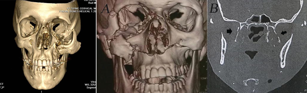
Common Facial Injuries Treated
Nasal Bone Fracture
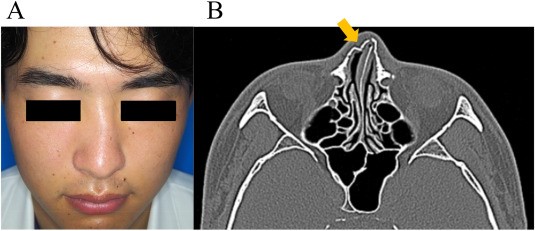
Common after falls, sports injuries, road traffic accidents, or assault.
Symptoms may include:
- Nasal deformity
- Swelling
- Nasal blockage
- Bleeding from nose
- Difficulty breathing
Mandible (Jaw) Fracture
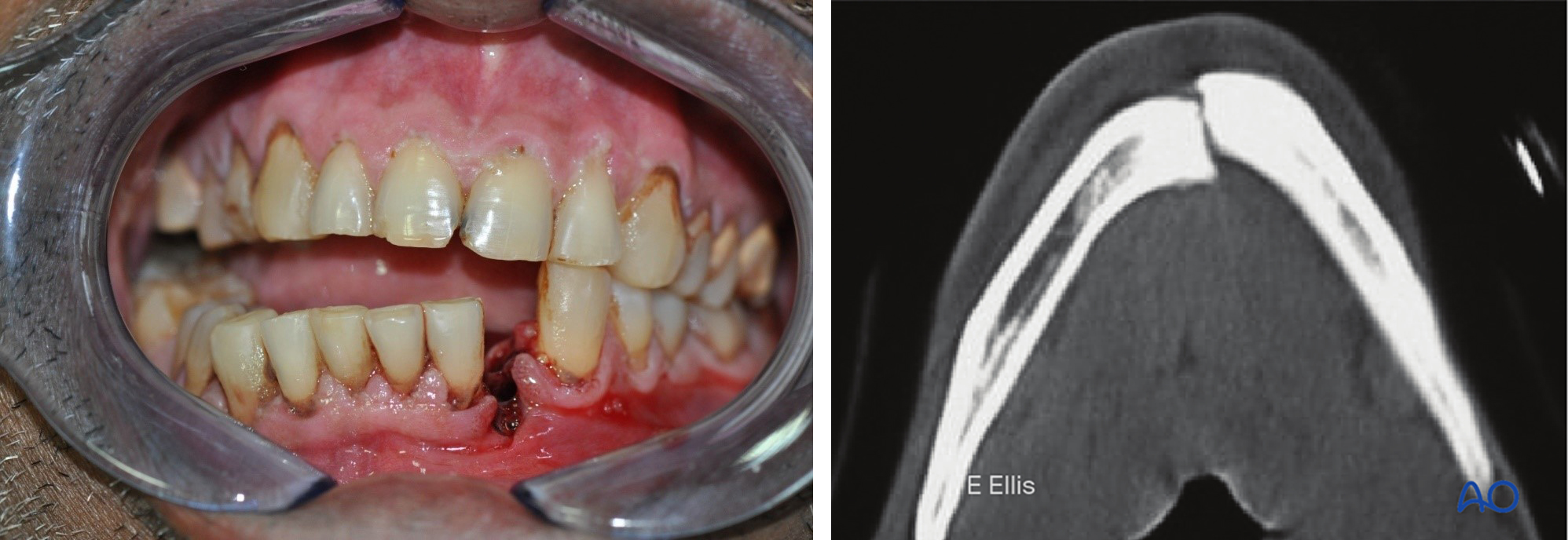
Can affect chewing, speech, and dental alignment.
Symptoms:
- Pain while opening mouth
- Misaligned bite
- Facial swelling
- Difficulty eating
- Loose teeth
Zygomatic (Cheek Bone) Fracture
May cause:
- Flattened cheek
- Facial asymmetry
- Double vision
- Difficulty opening mouth
Orbital (Eye Socket) Fracture
Can lead to:
- Swelling around eye
- Double vision
- Eye movement restriction
- Sunken appearance of eye
Maxillary Fractures
Upper jaw fractures affecting facial support and bite alignment.
Facial Lacerations
Cuts and soft tissue injuries requiring careful cosmetic repair.
Emergency Evaluation
Facial trauma assessment may include:
- Clinical examination
- CT scan of face
- Nasal endoscopy
- Eye examination
- Dental and bite assessment
Airway safety and associated head or neck injuries are always carefully evaluated.
Treatment Options
- Closed reduction: Used for selected nasal and facial fractures without major displacement.
- Open reduction and internal fixation (ORIF): Titanium plates and screws are used to stabilize facial bones in proper position.
- Endoscopic fracture management: Minimally invasive techniques may be used in selected cases.
- Soft tissue repair: Precise suturing techniques are used to minimize visible scarring.
Mandible (Jaw) Fracture Surgery
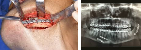
Jaw fractures may require:
- Plate fixation
- Arch bars
- Bite alignment correction
- Temporary dietary restriction
Goal of treatment:
- Restore normal bite
- Improve chewing
- Maintain facial symmetry
- Allow proper healing
Nasal Fracture Treatment
Nasal fractures are ideally treated within the first 1–2 weeks after injury before bones heal in displaced position.
Treatment may include:
- Closed reduction
- Septal hematoma drainage
- Septoplasty in selected cases
Recovery After Facial Trauma Surgery
Recovery depends on:
- Severity of injury
- Number of fractures
- Associated soft tissue damage
Common temporary symptoms:
- Swelling
- Bruising
- Mild numbness
- Jaw stiffness
- Nasal blockage
Most swelling improves significantly within 2–3 weeks.
After Surgery Instructions
- Avoid facial trauma or pressure.
- Eat soft diet if advised.
- Maintain oral hygiene carefully.
- Avoid smoking and alcohol.
- Use cold compresses initially.
- Take medications as prescribed.
When to Seek Urgent Care
Contact your doctor immediately if you develop:
- Difficulty breathing.
- Sudden vision changes.
- Severe bleeding.
- Fever.
- Increasing swelling.
- Bite misalignment.
- Persistent vomiting or severe headache.
Our Approach
We focus on:
- Functional recovery.
- Facial symmetry.
- Airway preservation.
- Minimal scarring.
- Early return to normal activity.
Modern surgical techniques help achieve excellent cosmetic and functional outcomes in most patients.
For emergency facial trauma consultation or surgical care, please contact our ENT clinic.
All these surgical procedures for facial trauma are covered under Mediclaim policy in cashless manner.
મેક્સિલોફેશિયલ ટ્રોમા સર્જરી (ચહેરા અને જડબાના ઇજાની સર્જરી)
ચહેરા પર થયેલી ઇજાઓમાં ચહેરાના હાડકાં, જડબું, નાક, આંખના પોલાણ (Eye Socket), ગાલના હાડકાં તેમજ નરમ પેશીઓને નુકસાન થઈ શકે છે.
મેક્સિલોફેશિયલ ટ્રોમા સર્જરીનો મુખ્ય હેતુ ઇજા પછી ચહેરાની રચના, કાર્યક્ષમતા, શ્વાસ લેવામાં સરળતા, બોલવાની ક્ષમતા, ચાવવામાં સરળતા અને કુદરતી દેખાવને પુનઃસ્થાપિત કરવાનો છે.
અમારા ENT વિભાગમાં આધુનિક ઇમેજિંગ, એન્ડોસ્કોપિક ટેક્નોલોજી અને અત્યંત ચોક્કસ ફ્રેક્ચર ફિક્સેશન પદ્ધતિઓ દ્વારા પસંદગીના ચહેરાના ટ્રોમા કેસોની સારવાર કરવામાં આવે છે.
ચહેરાની સામાન્ય ઇજાઓ
નાકના હાડકાનું ફ્રેક્ચર (Nasal Bone Fracture)
આ સામાન્ય રીતે નીચેના કારણોસર થાય છે:
- પડી જવું
- રમતગમત દરમિયાન ઇજા
- રોડ ટ્રાફિક અકસ્માત
- મારામારી અથવા અન્ય ઇજા
સામાન્ય લક્ષણો:
- નાક વાંકો થઈ જવો
- સોજો આવવો
- નાક બંધ થઈ જવું
- નાકમાંથી લોહી આવવું
- શ્વાસ લેવામાં તકલીફ પડવી
જડબાનું ફ્રેક્ચર (Mandible Fracture)
આ પ્રકારની ઇજા નીચેની બાબતોને અસર કરી શકે છે:
- ચાવવામાં તકલીફ
- બોલવામાં મુશ્કેલી
- દાંતની ગોઠવણી બગડવી
સામાન્ય લક્ષણો:
- મોઢું ખોલતી વખતે દુખાવો
- દાંતની ગોઠવણી બદલાઈ જવી
- ચહેરા પર સોજો
- ખોરાક ખાવામાં મુશ્કેલી
- દાંત હલી જવા
ગાલના હાડકાનું ફ્રેક્ચર (Zygomatic Fracture)
આમાં નીચે મુજબની સમસ્યાઓ થઈ શકે છે:
- ગાલ દબાયેલો દેખાવા લાગવો
- ચહેરાની અસમાનતા
- બેવડું દેખાવું (Double Vision)
- મોઢું ખોલવામાં મુશ્કેલી
આંખના પોલાણનું ફ્રેક્ચર (Orbital Fracture)
આમાં નીચે મુજબની સમસ્યાઓ થઈ શકે છે:
- આંખની આસપાસ સોજો
- બેવડું દેખાવું
- આંખ હલાવવામાં તકલીફ
- આંખ અંદર ધસી ગયેલી દેખાવવી
ઉપરના જડબાનું ફ્રેક્ચર (Maxillary Fracture)
આ પ્રકારના ફ્રેક્ચર ચહેરાના આધાર અને દાંતની ગોઠવણીને અસર કરે છે.
ચહેરાના કાપા અને ઇજાઓ (Facial Lacerations)
ચહેરા પર પડેલા કાપા અને નરમ પેશીઓની ઇજાઓ માટે સુંદર કોસ્મેટિક પરિણામ મળે તે રીતે અત્યંત કાળજીપૂર્વક ટાંકા લેવામાં આવે છે.
ઇમરજન્સી મૂલ્યાંકન
ચહેરાની ઇજાની તપાસમાં નીચે મુજબનો સમાવેશ થઈ શકે છે:
- સંપૂર્ણ ક્લિનિકલ તપાસ
- ચહેરાનો CT Scan
- નાકની એન્ડોસ્કોપી
- આંખોની તપાસ
- દાંત અને Bite ની તપાસ
દર્દીની શ્વાસનળી સુરક્ષિત છે કે નહીં તેમજ માથા અને ગરદનની અન્ય ગંભીર ઇજાઓ પણ હંમેશા તપાસવામાં આવે છે.
સારવારની પદ્ધતિઓ
- Closed Reduction: વધુ ખસેડાયેલા ન હોય એવા નાક અને ચહેરાના કેટલાક ફ્રેક્ચરમાં કરવામાં આવે છે.
- Open Reduction & Internal Fixation (ORIF): ટાઇટેનિયમ પ્લેટ અને સ્ક્રૂ દ્વારા હાડકાંને યોગ્ય સ્થિતિમાં મજબૂત રીતે સ્થિર કરવામાં આવે છે.
- એન્ડોસ્કોપિક ફ્રેક્ચર સર્જરી: પસંદગીના કેસોમાં ઓછી ચીરફાડવાળી એન્ડોસ્કોપિક ટેક્નિકનો ઉપયોગ કરવામાં આવે છે.
- Soft Tissue Repair: ચહેરા પર ઓછામાં ઓછો ડાઘ રહે તે માટે અત્યંત ચોક્કસ ટાંકા લેવામાં આવે છે.
જડબાના ફ્રેક્ચરની સર્જરી
જડબાના ફ્રેક્ચરમાં નીચે મુજબની સારવાર જરૂરી બની શકે છે:
- પ્લેટ ફિક્સેશન
- આર્ચ બાર્સ
- દાંતની ગોઠવણી સુધારવી
- અમુક સમય સુધી નરમ ખોરાક લેવો
સારવારનો મુખ્ય હેતુ:
- દાંતની સામાન્ય ગોઠવણી પાછી લાવવી
- યોગ્ય રીતે ચાવી શકાય તેવું બનાવવું
- ચહેરાની સમાનતા જાળવવી
- હાડકું યોગ્ય રીતે જોડાય તેની ખાતરી કરવી
નાકના ફ્રેક્ચરની સારવાર
નાકના હાડકાના ફ્રેક્ચરની સારવાર સામાન્ય રીતે ઇજા થયાના પ્રથમ ૧ થી ૨ અઠવાડિયામાં કરવી શ્રેષ્ઠ રહે છે, કારણ કે ત્યારબાદ હાડકાં ખસી ગયેલી સ્થિતિમાં જ જોડાવા લાગે છે.
સારવારમાં નીચે મુજબનો સમાવેશ થઈ શકે છે:
- ક્લોઝ્ડ રિડક્શન (Closed Reduction)
- સેપ્ટલ હેમેટોમા ડ્રેનેજ (Septal Hematoma Drainage)
- પસંદગીના દર્દીઓમાં સેપ્ટોપ્લાસ્ટી (Septoplasty)
ચહેરાની સર્જરી પછીની રિકવરી
સાજા થવાનો સમય નીચેની બાબતો પર આધાર રાખે છે:
- ઇજાની ગંભીરતા
- ફ્રેક્ચરની સંખ્યા
- નરમ પેશીઓને થયેલું નુકસાન
સર્જરી પછી સામાન્ય રીતે નીચે મુજબની તકલીફો થોડા સમય માટે થઈ શકે છે:
- સોજો
- વાદળી ડાઘ (Bruising)
- થોડી સુન્નતા (Numbness)
- જડબું જકડાઈ જવું
- નાક બંધ લાગવું
મોટાભાગનો સોજો સામાન્ય રીતે ૨ થી ૩ અઠવાડિયામાં નોંધપાત્ર રીતે ઓછો થઈ જાય છે.
સર્જરી પછીની મહત્વપૂર્ણ સૂચનાઓ
- ચહેરા પર ફરીથી ઇજા ન થાય તેનું ખાસ ધ્યાન રાખો.
- ડૉક્ટરની સલાહ મુજબ થોડા દિવસ નરમ ખોરાક લો.
- મોઢાની સ્વચ્છતા જાળવો.
- ધૂમ્રપાન અને આલ્કોહોલથી દૂર રહો.
- શરૂઆતના દિવસોમાં ઠંડા પાણીની પટ્ટી (Cold Compress) કરો.
- ડૉક્ટર દ્વારા સૂચવેલી તમામ દવાઓ નિયમિત લો.
તાત્કાલિક ડૉક્ટરનો સંપર્ક ક્યારે કરવો?
જો નીચે મુજબની કોઈપણ તકલીફ થાય તો તરત જ ડૉક્ટરનો સંપર્ક કરો:
- શ્વાસ લેવામાં તકલીફ
- અચાનક આંખની દ્રષ્ટિમાં ફેરફાર
- વધુ પડતું લોહી નીકળવું
- તાવ આવવો
- સોજો સતત વધતો જવો
- દાંતની ગોઠવણી બગડી જવી
- સતત ઉલ્ટી અથવા અસહ્ય માથાનો દુખાવો
અમારો અભિગમ
અમારું મુખ્ય ધ્યાન નીચેની બાબતો પર રહે છે:
- ચહેરાની સંપૂર્ણ કાર્યક્ષમતા પાછી લાવવી.
- ચહેરાની કુદરતી સમાનતા જાળવવી.
- શ્વાસનળીને સુરક્ષિત રાખવી.
- ચહેરા પર ઓછામાં ઓછા ડાઘ રહે તે સુનિશ્ચિત કરવું.
- દર્દી ઝડપથી સામાન્ય જીવનમાં પાછા ફરી શકે તે માટે પ્રયત્ન કરવો.
આધુનિક સર્જિકલ ટેક્નોલોજીના કારણે મોટાભાગના દર્દીઓમાં કાર્યક્ષમતા અને કોસ્મેટિક પરિણામ બંને ખૂબ જ ઉત્તમ મળે છે.
ચહેરાની ઇજાના ઇમરજન્સી નિદાન અથવા સર્જિકલ સારવાર માટે કૃપા કરીને અમારા ENT વિભાગનો સંપર્ક કરો.
ચહેરાના ટ્રોમા માટે કરવામાં આવતી આ તમામ સર્જરીઓ મોટાભાગની Mediclaim પોલિસી હેઠળ Cashless સુવિધામાં ઉપલબ્ધ છે.
Departments
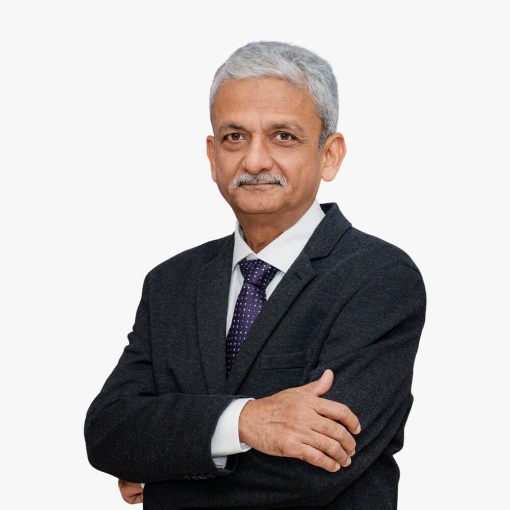
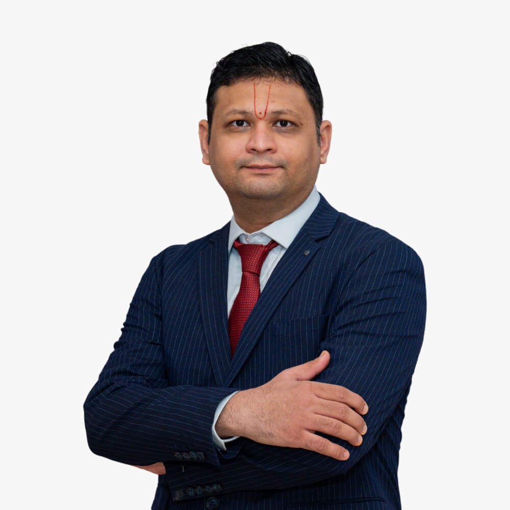
Dr. Nilay S. Shah
MS (ENT), ENT & Head - Neck Surgeon, Endoscopic Sinus, Snoring & Sleep Apnea Surgeon, Allergy & Vertigo Specialist.